Chapter 48
Chapter 48. Nervous Systems. Nerves with giant axons. Ganglia. Brain. Arm. Eye. Mantle. Nerve. Many animals have a complex nervous system which consists of: A central nervous system (CNS) where integration takes place; this includes the brain and a nerve cord

Chapter 48
E N D
Presentation Transcript
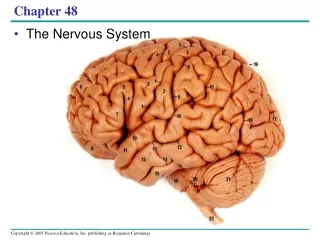
Chapter 48 Nervous Systems
Nerves with giant axons Ganglia Brain Arm Eye Mantle Nerve
Many animals have a complex nervous system which consists of: • A central nervous system (CNS) where integration takes place; this includes the brain and a nerve cord • A peripheral nervous system (PNS), which brings information into and out of the CNS
Sensory input Integration Sensor Motor output Central nervous system (CNS) Effector Peripheral nervous system (PNS)
Neuron Structure and Function • Neurons- nerve cells that transfer information within the body • Cell body- location of most of a neuron’s organelles • Dendrites- highly branched extensions that receive signals from other neurons • Axon- much longer extension that transmits signals to other cells at synapses
Neuron Structure and Function • Synapse- is a junction between an axon and another cell • Synaptic terminal- one axon passes information across the synapse in the form of chemical messengers called neurotransmitters • Glia- cells that nourish or insulate neurons
Dendrites Stimulus Presynaptic cell Nucleus Axon hillock Cell body Axon Synapse Synaptic terminals Postsynaptic cell Neurotransmitter
Ion pumps and ion channels maintain the resting potential of a neuron • Every cell has a voltage (difference in electrical charge) across its plasma membrane called a membrane potential • Messages are transmitted as changes in membrane potential
Resting Potential- the membrane potential of a neuron not sending signals • Concentration of K+ is greater inside the cell, while the concentration of Na+ is greater outside the cell • Sodium-potassium pumps use the energy of ATP to maintain these K+ and Na+ gradients • Ion channels- converts chemical potential to electrical potential • A neuron at resting potential contains many open K+ channels and fewer open Na+ channels; K+ diffuses out of the cell Animation: Resting Potential
Key Sodium- potassium pump Na+ Potassium channel Sodium channel K+ OUTSIDE CELL INSIDE CELL (b)
Action potential +50 Falling phase 0 Rising phase Membrane potential (mV) Threshold (–55) –50 Resting potential –70 Depolarization Undershoot –100 Time (msec)
An action potential occurs if a stimulus causes the membrane voltage to cross a particular threshold • An action potential is a brief all-or-none depolarization of a neuron’s plasma membrane • Action potentials are signals that carry information along axons
Generation of Action Potentials • At resting potential • 1. Most voltage-gated Na+ and K+ channels are closed, but some K+ channels (not voltage-gated) are open • When an action potential is generated • 2. Voltage-gated Na+ channels open first and Na+ flows into the cell • 3. During the rising phase, the membrane potential increases • 4. During the falling phase, voltage-gated Na+ channels become inactivated; voltage-gated K+ channels open, and K+ flows out of the cell • 5. During the undershoot, membrane permeability to K+ is at first higher than at rest, then voltage-gated K+ channels close; resting potential is restored
Key Na+ K+ Falling phase of the action potential 4 Rising phase of the action potential 3 +50 Action potential 3 0 Membrane potential (mV) 2 4 Threshold –50 1 1 5 Resting potential Depolarization 2 –100 Time Extracellular fluid Sodium channel Potassium channel Plasma membrane Cytosol Inactivation loop Undershoot 5 Resting state 1
Refractory period after an action potential, a second action potential cannot be initiated. • The refractory period is a result of a temporary inactivation of the Na+ channels BioFlix: How Neurons Work Animation: Action Potential
Axon Plasma membrane Action potential Cytosol Na+ Action potential K+ Na+ K+ Action potential K+ Na+ K+
Conduction Speed • Axons are insulated by a myelin sheath, which causes an action potential’s speed to increase • Myelin sheaths are made by glia— oligodendrocytesin the CNS and Schwann cells in the PNS
Node of Ranvier Layers of myelin Axon Schwann cell Schwann cell Nodes of Ranvier Nucleus of Schwann cell Axon Myelin sheath
Neurons communicate with other cells at synapses • At electrical synapses, the electrical current flows from one neuron to another • At chemical synapses, a chemical neurotransmitter carries information across the gap junction • Most synapses are chemical synapses Animation: Synapse
5 Na+ K+ Synaptic vesicles containing neurotransmitter Presynaptic membrane Voltage-gated Ca2+ channel Postsynaptic membrane Ca2+ 1 4 6 2 3 Synaptic cleft Ligand-gated ion channels
Neurotransmitters • The same neurotransmitter can produce different effects in different types of cells • There are five major classes of neurotransmitters: acetylcholine, biogenic amines, amino acids, neuropeptides, and gases
Acetylcholine- common neurotransmitter, usually an excitatory transmitter Biogenic Amines- include epinephrine, norepinephrine, dopamine, and serotonin
Amino Acid- gamma-aminobutyric acid (GABA) and glutamate Neuropeptides- relatively short chains of amino acids, include substance P and endorphins Gases- nitric oxide and carbon monoxide are local regulators in the PNS
Central nervous system (CNS) where integration takes place (brain and nerve cord) • Peripheral nervous system (PNS) brings information into and out of the CNS
Nervous systems consist of circuits of neurons and supporting cells • Nerve net- a series of interconnected nerve cells • Nerves- bundles that consist of the axons of multiple nerve cells
Radial nerve Nerve ring Nerve net (a) Hydra (cnidarian) (b) Sea star (echinoderm)
Eyespot Brain Brain Nerve cords Ventral nerve cord Transverse nerve Segmental ganglia (c) Planarian (flatworm) (d) Leech (annelid)
Brain Ganglia Anterior nerve ring Ventral nerve cord Longitudinal nerve cords Segmental ganglia (e) Insect (arthropod) (f) Chiton (mollusc)
Brain Spinal cord (dorsal nerve cord) Brain Sensory ganglia Ganglia (g) Squid (mollusc) (h) Salamander (vertebrate)
Organization of the Vertebrate Nervous System • Central nervous system (CNS) where integration takes place (brain and nerve cord) • Peripheral nervous system (PNS) brings information into and out of the CNS
Peripheral nervous system (PNS) Central nervous system (CNS) Brain Cranial nerves Spinal cord Ganglia outside CNS Spinal nerves
The brain and spinal cord contain • Gray matter- neuron cell bodies, dendrites, and unmyelinated axons • White matter- bundles of myelinated axons Gray matter White matter Ventricles
Cell body of sensory neuron in dorsal root ganglion Gray matter Quadriceps muscle White matter Hamstring muscle Spinal cord (cross section) Sensory neuron Motor neuron Interneuron
The Peripheral Nervous System • Afferent neurons transmit information to the CNS and efferent neurons transmit information away from the CNS • Motor system- carries signals to skeletal muscles and is voluntary • Autonomic nervous system- regulates the internal environment involuntarily • Sympathetic division- correlates with the “fight-or-flight” response • Parasympathetic division- promotes a return to “rest and digest” • Enteric division- controls activity of the digestive tract, pancreas, and gallbladder
PNS Afferent (sensory) neurons Efferent neurons Autonomic nervous system Motor system Hearing Sympathetic division Parasympathetic division Enteric division Locomotion Hormone action Gas exchange Circulation Digestion
Sympathetic division Parasympathetic division Action on target organs: Action on target organs: Dilates pupil of eye Constricts pupil of eye Inhibits salivary gland secretion Stimulates salivary gland secretion Sympathetic ganglia Constricts bronchi in lungs Relaxes bronchi in lungs Cervical Slows heart Accelerates heart Stimulates activity of stomach and intestines Inhibits activity of stomach and intestines Thoracic Stimulates activity of pancreas Inhibits activity of pancreas Stimulates glucose release from liver; inhibits gallbladder Stimulates gallbladder Lumbar Stimulates adrenal medulla Promotes emptying of bladder Inhibits emptying of bladder Sacral Promotes erection of genitals Promotes ejaculation and vaginal contractions Synapse
Cerebrum (includes cerebral cortex, white matter, basal nuclei) Telencephalon Forebrain Diencephalon Diencephalon (thalamus, hypothalamus, epithalamus) Midbrain Mesencephalon Midbrain (part of brainstem) Metencephalon Pons (part of brainstem), cerebellum Hindbrain Myelencephalon Medulla oblongata (part of brainstem) Diencephalon: Cerebrum Mesencephalon Hypothalamus Metencephalon Thalamus Midbrain Pineal gland (part of epithalamus) Myelencephalon Hindbrain Diencephalon Brainstem: Midbrain Pons Spinal cord Pituitary gland Forebrain Medulla oblongata Telencephalon Spinal cord Cerebellum Central canal (c) Adult (a) Embryo at 1 month (b) Embryo at 5 weeks
Cerebral cortex Cerebrum Thalamus Forebrain Hypothalamus Pituitary gland Midbrain Pons Spinal cord Medulla oblongata Hindbrain Cerebellum
The Brainstem • Brainstem- coordinates and conducts information between brain centers • Midbrain- contains centers for receipt and integration of sensory information • Pons- regulates breathing centers in the medulla • Medulla oblongata- controls breathing, cardiovascular activity, swallowing, vomiting, and digestion The brainstem and cerebrum control arousal and sleep
The Cerebellum • Cerebellum- coordination and error checking during motor, perceptual, and cognitive functions • It is also involved in learning and remembering motor skills
The Cerebrum • Corpus callosum- a thick band of axons provides communication between the right and left cerebral cortices • The right half of the cerebral cortex controls the left side of the body, and vice versa • Left hemisphere- language, math, logic, and processing of serial sequences • Right hemisphere- pattern recognition, nonverbal thinking, and emotional processing
Right cerebral hemisphere Left cerebral hemisphere Thalamus Corpus callosum Basal nuclei Cerebral cortex
Cerebral cortex- controls voluntary movement and cognitive functions • Each side of the cerebral cortex has four lobes: frontal, temporal, occipital, and parietal
Frontal lobe Parietal lobe Somatosensory cortex Motor cortex Somatosensory association area Speech Frontal association area Taste Reading Speech Hearing Visual association area Smell Auditory association area Vision Temporal lobe Occipital lobe
Max Seeing words Hearing words Min Speaking words Generating words
Memory and Learning • Learning can occur when neurons make new connections or when the strength of existing neural connections changes • Short-term memory is accessed via the hippocampus • The hippocampus also plays a role in forming long-term memory, which is stored in the cerebral cortex
Fig. 49-18 Thalamus Hypothalamus Prefrontal cortex Olfactory bulb Hippocampus Amygdala
Stem Cell–Based Therapy • The adult human brain contains stem cells that can differentiate into mature neurons