Download
1 / 17
351 likes | 1.11k Views
Chapter 19: Cardiovascular System: Blood Vessels. Étienne-Jules Marey – french physiologist born in 1830 credited with the invention of the sphymanograph (which has evolved into the sphymanometer (blood pressure cuff) used today.

E N D
Chapter 19: Cardiovascular System: Blood Vessels
Étienne-Jules Marey – french physiologist born in 1830 credited with the invention of the sphymanograph (which has evolved into the sphymanometer (blood pressure cuff) used today.
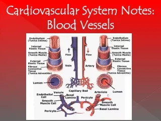
Figure 19.1: Generalized structure of arteries, veins, and capillaries, p. 715. Artery Vein (a) Valve Tunica intima • Endothelium • Subendothelial layer Internal elastic lamina Tunica media External elastic lamina Tunica externa Lumen Lumen Capillary network Vein Artery Endothelial cells (b) Capillary
Figure 19.2: Overview of vascular components and blood distribution, p. 717. Venous system Arterial system Heart Large veins Elastic arteries (conducting vessels) Large lymphatic vessels Capacitance vessels Lymph node Muscular arteries (distributing vessels) Lymphatic system Small veins Arteriovenous anastomosis Lymphatic capillary Arterioles (resistance vessels) Postcapillary venule Terminal arteriole Sinusoid Metarteriole Precapillary sphincter Thoroughfare channel Pulmonary blood vessels 12% (a) Capillaries (exchange vessels) Heart 8% Systemic arteries and arterioles 15% Systemic veins and venules 60% (b) Capillaries 5%
Figure 19.4: Anatomy of a capillary bed, p. 721. Vascular shunt Precapillary sphincters Metarteriole Thoroughfare channel True capillaries Terminal arteriole Postcapillary venule (a) Sphincters open Terminal arteriole Postcapillary venule (b) Sphincters closed
Figure 19.5: Blood pressure in various blood vessels of the systemic circulation, p. 725. 120 Systolic pressure 100 Mean pressure 80 Blood pressure (mm Hg) 60 Diastolic pressure 40 20 0 Veins Aorta Venules Arteries Arterioles Capillaries Venae cavae
Figure 19.3: Capillary structure, p. 720. Pericyte Pericyte Pinocytotic vesicles Red blood cell in lumen Red blood cell in lumen Intercellular cleft Endothelial cell Fenestra- tions (pores) Basement membrane Intercellular cleft Endothelial nucleus Tight junction Pinocytotic vesicles Endothelial nucleus Basement membrane Endothelialcell Tight junction (b) (a) Pericyte Endothelial cell Red blood cell in lumen Large intercellular cleft Tight junction Incomplete basement membrane Nucleus of endothelial cell (c)
Figure 19.6: The muscular pump, p. 726. Valve (open) Contracted skeletal muscle Valve (closed) Vein Direction of blood flow
Figure 19.8a: Baroreceptor reflexes that help maintain blood pressure homeostasis, p. 728. Impulse traveling along afferent nerves from baroreceptors: Stimulate cardio- inhibitory center (and inhibit cardio- acceleratory center) Sympathetic impulses to heart ( HR and contractility) Baroreceptors in carotid sinuses and aortic arch stimulated CO Inhibit vasomotor center R Arterial blood pressure rises above normal range Rate of vasomotor impulses allows vasodilation ( vessel diameter) CO and R return blood pressure to homeostatic range Stimulus: Rising blood pressure Imbalance Homeostasis: Blood pressure in normal range Imbalance
Figure 19.8b: Baroreceptor reflexes that help maintain blood pressure homeostasis, p. 728. Imbalance Homeostasis: Blood pressure in normal range Stimulus: Declining blood pressure Imbalance CO and R return blood pressure to Homeostatic range Impulses from baroreceptors: Stimulate cardio- acceleratory center (and inhibit cardio- inhibitory center) Arterial blood pressure falls below normal range Cardiac output (CO) Baroreceptors in carotid sinuses and aortic arch inhibited Sympathetic impulses to heart ( HR and contractility) Peripheral resistance (R) Vasomotor fibers stimulate vasoconstriction Stimulate vasomotor center
Figure 19.11: Body sites where the pulse is most easily palpated, p. 732. Temporal artery Facial artery Common carotid artery Brachial artery Radial artery Femoral artery Popliteal artery Posterior tibial artery Dorsalis pedis artery
Figure 19.18a: Pulmonary circulation, p. 744. Pulmonary capillaries of the R. lung Pulmonary capillaries of the L. lung R. pulmonary artery L. pulmonary artery To systemic circulation Pulmonary trunk R. pulmonary veins From systemic circulation RA LA L. pulmonary veins RV LV (a)
Figure 19.18b: Pulmonary circulation, p. 744. Left pulmonary artery Air-filled alveolus of lung Aortic arch Pulmonary trunk Right pulmonary artery O2 Three lobar arteries to right lung CO2 Pulmonary capillary Gas exchange Pulmonary veins Two lobar arteries to left lung Right atrium Pulmonary veins Left atrium Right ventricle Left ventricle (b)