CLASSICAL GLENN
Fig 55. CLASSICAL GLENN. BIDIRECTIONAL GLENN. FONTAN. SVC. SVC. RPA. LPA. PA. PA. RA. RA. RA. IVC. IVC. TOTAL CAVOPULMONARY CONNECTION. SVC. PA. RA. IVC. Fig 56. Tricuspid atresia. Apical four chamber view of a univentricular atrioventricular connection with atresia of the
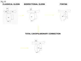

CLASSICAL GLENN
E N D
Presentation Transcript
Fig 55 CLASSICAL GLENN BIDIRECTIONAL GLENN FONTAN SVC SVC RPA LPA PA PA RA RA RA IVC IVC TOTAL CAVOPULMONARY CONNECTION SVC PA RA IVC
Fig 56 Tricuspid atresia Apical four chamber view of a univentricular atrioventricular connection with atresia of the right sided atriovenricular valve (tricuspid atresia).The patient had a Fontan type repair (right atrium to pulmonary artery connection). The valve between the IVC and right atrium can just be seen in the right atrium (arrow). MLV LA RA
Fig 57 TCPC Apical four chamber view of a patient with a univentricular atriovenricular connection and a common atrium, post TCPC. Note the TCPC pathway (arrow) at the back of the atrium. V Common atrium
Fig 58 Tricuspid atresia post Fontan operation Apical view of a univentricular atrioventricular connection resulting from atresia of the right-sided atrioventricular valve (tricuspid atresia). The patient had a Fontan type operation. Note that the right atrium is severely dilated with thrombus inside. LV LA RA
Fig 59 Fontan: The giant RA and large clot RA RA TOE image taken from a patient with a double inlet left ventricle post Fontan (atria-pulmonary connection) repair. Note the severely dilated right atrium with giant thrombus and spontaneous contrast inside the atrial chamber (arrow).