Download

1 / 61
620 likes | 801 Views
Chapter 4. Viruses —— Non-cellular entities. Section A. Introduction A-1. Definition Contain only a single type of nucleic acid, either DNA or RNA.

E N D
Chapter 4. Viruses —— Non-cellular entities
Section A. Introduction A-1. Definition Contain only a single type of nucleic acid, either DNA or RNA. Contain a protein coat (sometimes itself enclosed by an envelope of lipids, protein, and carbohydrates) that surrounds the nucleic acid. Multiply inside living cells by using the synthesizing machinery of the cell. They are simple, acellular entities, may be regarded as an exceptionally complex molecular microorganism .
A-2. Development of Virology • Although the ancients did not understand the nature of illnesses, such as rabies, smallpox, they were acquainted with disease that are now known to be viral in origin. • In 1886,the Dutch chemist Adolf Mayer (German) showed that tobacco mosaic disease was transmissible. In 1892, the Russian bacteriologist Dimitri Iwanowski proved that the infectious agent of the disease could pass through the porcelain filter and contracted tobacco mosaic disease. In 1898, the Dutch botanist Martinus Beijeringck observed that the behavior of this agent was caused by a filterable virus (Latin, for poison) ,which would multiply only in living plant cell (1890).
The Russian microbiologist Winograsky discovered that soil bacteria could oxidize iron, sulfur and ammonia to obtain energy, and also isolated nitrogen – fixing bacteria. Beijerinck made fundamental contributions to microbial ecology. He isolated Azotobacter and Rhizobium.
At the same time, many other diseases were found to be caused by the filterable virus by other scientists. The term virus have been commonly used up to now. • In 1935, American chemist Wendell M. Stainley (of the Rockefeller Institute) isolated the tobacco mosaic virus and successfully crystallized the virus, making it possible for the first time to carry out chemical and structural studies on a purified virus. Stainley was later awarded the 1946 Nobel Prize in chemistry. • At about the same time (early 30s), the invention of electron microscope made it possible to see viruses. Kausche (German, 1940) observed the rod-like shape of TMV for the first time.
From 1950s, molecular biology of virus had made a great progresses in the feature, components, structure, even the sequencing of viral genomes. • Advances in molecular biology techniques have led to the recognition of new human viruses. Newly recognized viruses are referred to as emerging viruses-viruses that are not necessarily new but that may be new to Western medicine. • HIV (human immunodeficiency virus) was recognized as the agent of AIDS (acquired immunodeficiency syndrome) in 1983. EHF (Ebola hemorrahgic fever) have been learned in Zaire in 1995. And the virus (coronavirus)responsible for the SARS (severe acute respiratory syndrome) as you know in 2003.
HIV INFLU
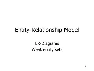
Orf virus:口疮病毒(接触性脓疱皮炎病毒) (负染技术) Vaccinia virus痘苗病毒 (投影技术) Ebola virus :埃博拉病毒(正染技术)
Section B. Virus B-1. Host Range The host range of a virus is the spectrum of host cells the virus can infect. How ever, most viruses are able to infect specific types of only one host species. bacterial phage phycophage mycophage Phage Animal viruses Plant virus Protozoal viruses invertebrate viruses vertebrate viruses Viruses
B-2. Viral Size Viral sizes are determined with the aid of electron microscopy. Different viruses vary considerably in size. Although most are quite a bit smaller than bacteria, some of the larger viruses (such as the vaccinia virus or poxviruses) are about the same size as some very small bacteria (such as mycoplasmas). Viruse range from 20 to 14,000 nm in length. Largest: Poxviruses 450 nm Longest: Filovirus 80-14,000 nm Smallest: Bean distortion dwarf Thinnest: Coliphage f1 5 × 800 nm (Ebola virus) virus (BBDV) 9-11
B-3. Viral Structure 3-1. Virion A virion is a complete, fully developed viral particle composed of nucleic acid and surrounded by a protein coat called Capsid that protects it from the environment and is a vehicle of transmission from one host cell to another. Capsid made up of protomers (capsomer) Genome (core): DNA or RNA Envelope Virion
3-2. Symmetries and Morphology Viruses may be classified in to tree basic different morphological types based on their capsid architecture. The structure of these capsids has been revealed by electron microscopy and a technique called X-ray crystallography. 3-2.1 Helical symmetry Helical capsids are shaped much like hollow tubes with protein walls. TMV provides a well-studied example of helical capsid structure. RNA is wound in a spiral and positioned toward the inside of the capsid where it lies within a groove formed by the protomers (capsomers or protein particles).
Helical symmetry Icosahedral symmetry Complex symmetry
3-2.2 Icosahedral symmetry or Polyhedral symmetry The icosahedron is one of nature ’ s favorite shape.( the helix is probably most popular) Viruses employ the icosahedral shape because it’s the most efficient way to enclose a space. A few genes, sometimes only one, can code for proteins that self-assemble to form the capsid. In this way, a small number of linear genes can specify a large three-dimensional structure. The capsids are constructed from ring-or knob-shaped units called capsomers or protomers, they usually form a unit of five or six protomers named pentons and hexons which form the 12 corners (apexex) and 20 triangular faces. N=10 ( n-1 )2 +2
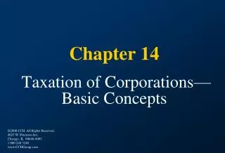
3-2.3 Complex symmetry Some viruses do not fit into either the category. The poxviruses and T-even coliphages are two important examples. They are complex symmetries with both heads (icosahedral) and tails (helical) combined together (such as T2 , T4 , T6) head collar Core or tube Helical sheath Fiber Base plate pins T-2 coliphage
3-2.4 Enveloped viruses Many animal viruses, some plant viruses are bounded by an outer membranous layer called an envelope. Animal viral envelopes usually arise from host cell nuclear or plasma membranes; their lipids and carbohydrates are normal host constituents. In contrast, envelope proteins are coded by virus genes and may even project from the envelope surface as spikes or peplomers . Influenza virus is a well-studied example of an enveloped virus. 3-3 Chemical Compositions Structural (protomer)functional (enzymes) ssDNA dsDNA ssRNA dsRNA proteins DNA RNA Nucleic acid +RNA -RNA
Functional proteins are of non-structural proteins mostly exist freely from the structures and functioned as enzymes, or the infectious factors. Such as attachment or penetrating associate glycoprotein spikes on the surface of HIV, influenza viruses, or the tail fibers of bacterial phages. Certain viruses contain enzymes of neuraminidase responsible to the damage of host plasma membrane or cell wall. Most functional proteins found in viruses are duplication associate enzymes, such as polymerases of DNA or RNA, reverse transcriptase, also the protein synthesis associated enzymes.
Other components Other components refer mainly to the substances found in envelopes of complexity virions. Lipids and carbohydrates are often found within the envelopes that usually arise from host cell nuclear or plasma membranes. That is why they resemble the membrane components and structures. Carbohydrates exist generally in forms of glycoprotein and responsible for their antigenicities.
B-4. Replication or Multiplication Although the means by which a virus enters and exits a host cell may vary, the basic mechanism of viral multiplication is similar for all viruses. The best understood viral life cycles are those of the bacteriophage. So that we will take the T-even coliphages as an example of the life (lytic) cycle. The multiplication (life) cycle of phages, like that of all viruses, occurs in five distinct stages: attachment (adsorption) penetration biosynthesis assembly (maturation) release or burst
4-1. Attachment After a chance collision between phage particles and bacteria, attachment or adsorption, occurs. During this process, an attachment site on the virus attaches to a complementary receptor site on the bacterial cell. This attachment is a chemical interaction in which weak bonds are formed between the attachment and receptor sites. T-even coliphages use fibers at the end of the tail as attachment sites. The complementary receptor sites are on the bacterial cell wall. This gives the specific feature of adsorption. The receptors and the attachment sites are the chemical fundamentals of the specific adsorption. All the viruses and hosts have a similar mechanism.
4-2. Penetration After attachment, the phage fixed onto the surface of the host cell, and a stimulation was given. Then the tail sheath would contract to be half of its length, and the tail core (tube) insert into the host cell wall and the membrane. This process needs the help of ATPase and phage lysozyme. They are carried within the tube and helpful to the ATP release and the peptidoglycan digestion (degradation). After that, the DNA from the phage head,pass through the tail core (tube), and enters (be injected into) the cell. The capsid of the phage remains outside of the cell wall.
4-3. Biosynthesis (Replication) Once the phage DNA has reached the cytoplasm of the host cell, the biosynthesis of viral nucleic acid and protein occurs. Host protein synthesis is stopped by virus-induced degradation of the host DNA, viral proteins that interfere with transcription or the repression of translation. Genetic controls regulate when different regions of phage DNA are transcribed into mRNA during the multiplication cycle.
Since the viruses contain different genetic material (DNA or RNA) as their genetic information depend on the species, the modes of mRNA transcription are different from each other. There are six patterns found in viral mRNA transcription: ssDNA (±DNA) + DNA -DNA mRNA dsDNA (± DNA) -DNA mRNA dsRNA ( ± RNA) -RNA mRNA ssRNA ± RNA -RNA mRNA ± DNA +DNA - DNA mRNA +RNA - RNA mRNA
4-4. Assembly (Maturation) In the next sequence of events, maturation (assembly) occurs. In this process, the phage DNA and capsids are assembled into complete virions. The viral component essentially assembled into a viral particle spontaneously, eliminating the need for many non-structural genes and gene products. The phage heads and tails are separately assembled from protein subunits, and the head is filled with phage DNA and attached to the tail. 4-5. Release The final stage of multiplication is the release of virions from the host cell. The term lysis is generally used for this stage in T-even phages because of the cell membrane actually broken up and releasing virions.
B-5. One-Step Growth Curve One-Step growth curve is a classical experiment designed for demonstrating the multiplication of phages. In this procedure, a phage suspension is diluted until a sample containing only a few phage particles is obtained. These particles are then introduced into a culture of host cells. Periodically, samples of phage particles are removed from the culture and inoculated onto a plate culture of susceptible host cells; the plaque method is used to determine the number of infective phage particles on this culture. The curve drawn out contains three important stages: Latent phase; Burst (rise phase); and plateau. The number of phages newly released from a single cell is referred as to Burst Size (50-200)
One-step growth curve was designed by Max Delbrük and Emory Ellis in 1939. It is significant to the understanding of bacteriophages with three important aspects: The Latent phase; Burst (rise phase) and Burst size. This experiment is a remarkable event which starts the beginning of modern virology. And it is also an evidence of becteriophage reproduction mode of mutiplication, other than fission.
Latent phase: is the period from the infection to the completely formation of the first phage. There is no release of virions. At the very beginning of this phase, host cell contains no complete, infectious viral particles. This phenomenon can be observed by lysing the cells with chloroform. The initial period is called eclipse period. Burst (or rise) phase: This is the period that closely following the latent phase. In this rise phase, the bacterial cells lyse rapidly and release mature, infectious virions. Since the host cells are not synchronized individually, the burst of cells last for a period, other than a theoretically sudden burst. Plateau phase: This period is the final stage that the plaque is formed and no more viruses are released. The total number of virions released per cell is called burst size.
B-6. Lysogeny Some phages( viruses) do not cause lysis and death of the host cell when they infect and multiply.( such as coliphage λ ) They can either proceed through alytic cycle (in some certain conditions) or incorporate (integrate) their DNA into the host DNA. In the latter state, called Lysogeny, the phage remains latent (inactive). The phages that certainly cause lysis and death of the host cells are called Virulent Phage. The phages that can integrate their DNA into the host DNA and usually do not cause lysis of the cells are named Temperate Phage.