Download
1 / 16
160 likes | 413 Views
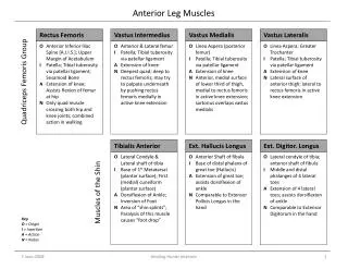
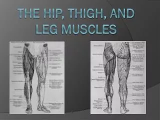
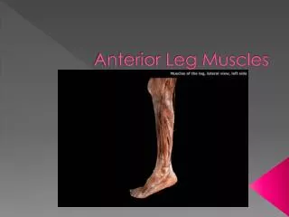
Anterior Leg Muscles. Tibialis Anterior. Origin: 1. lateral tibial condyle 2. proximal 2/3s of anterolateral surface of Tibia 3. Interosseous membrane, anterior intermuscular septum & cural fascia Insertion: 1. medial & plantar surface of base of 1 st metatarsal

E N D
Tibialis Anterior • Origin: 1. lateral tibial condyle 2. proximal 2/3s of anterolateral surface of Tibia 3. Interosseous membrane, anterior intermuscular septum & cural fascia • Insertion: 1. medial & plantar surface of base of 1st metatarsal 2. medial & plantar surface of the cuneiform • Action: powerfully dorsiflexes the foot, and inverts and adducts the foot! • Blood supply: anterior tibial artery • Nervous innervation: deep peroneal nerve , L4, S1
Extensor Hallucis longus • Origin: 1. medial aspect of the fibula 2. interosseous membrane, cural fascia • Insertion: 1. dorsal surface of base of proximal and distal phalanx of hallux • Action: 1. extends the distal phalanx of the big toe 2. weakly dorsiflexes the foot 3. weakly inverts & adducts the foot Blood: Anterior tibial artery Nervous Innervation: Deep peroneal nerve, L4, S1
Extensor digitorum longus • Origin: 1. upper anterior surface of fibula 2. interosseous membrane, crural fascia 3. lateral condyle of the tibia • Insertion: dorsal surface of the bases of the middle & distal phalanxes of the 2-5h rays (via 4 tendons and fibrous expansion) • Action: 1. extends the lateral 4 toes 2. weakly dorsiflexes & everts the foot • Blood: anterior tibial artery • Nervous innervation: deep peroneal nerve, L4, S1
Peroneus tertius • Origin: 1. distal 1/3 of anterior fibula 2. distal & lateral aspect of extensor digitorum • Insertion: doral surface of base of 5th metatarsal • Action: 1. extend the 5th metatarsal 2. weakly dorsiflexes & everts the foot • Blood: Anterior tibial artery • Nervous innervation: deep peroneal nerve, L4, S1
Quadratus plante • Origin: medial head: medial calcaneus lateral head: lateral calcaneus & long plantar ligament • Insertion: 1. lateral margin of tendon of flexor digitorum longus 2. may send slips into the distal tendons • Action: 1. assists FDL to flex the distal phalanxes of the 2-5 toes 2. corrects FDLfrom pulling toes medially • Blood: lateral plantar artery • Nervous innervation: lateral plantar nerver, S1&2