Download

1 / 13
130 likes | 366 Views
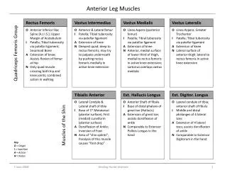
Deep Structures of the Anterior Leg. Christine Grecco & Tobias Johnson. Deep Muscles. Muscles Obturator externus Adductor magnus Adductor minimus Gracilis Adductor hiatus Vastus medialis. Deep muscles. Muscles Vastus intermedius Rectus femoris Sartorius Gracilis

E N D
Deep Structures of theAnterior Leg Christine Grecco & Tobias Johnson
Deep Muscles • Muscles • Obturatorexternus • Adductor magnus • Adductor minimus • Gracilis • Adductor hiatus • Vastusmedialis
Deep muscles • Muscles • Vastusintermedius • Rectus femoris • Sartorius • Gracilis • Adductor magnus • Vastusmedialis
Tendons • Tendons • Pesanserinus • Sartorius • Semitendinosus • Adductor hiatus • Adductor magnus • Gracilis
Deep Muscles & Tendons • Muscles • Vastusmedialis • Tendons • Adductor Magnus tendon • Gastrocnemius tendon, medial & lateral head
Bursa • Suprapatellar bursa • Superficial prepatellar burse • Deep infrapatellar bursa • Superficial infrapatellar bursa • Pes anserine bursa • Subsartorial bursa • Iliopectineal bursa (not pictured)
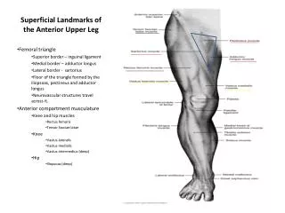
Femoral vessels Tracing the supply of the lower extremeties • Common Femoral Artery (CFA) • Travels two-thirds of the thigh • Pierces the adductor magnus • Reaches the poplitealfossa behind the knee • Lies on the psoas major, pectineus • Branches to the deep (DFA) and superficial femoral artery CFA DFA
Femoral vessels Tracing the supply of the lower extremities • Deep Femoral Artery (DFA) • Also known as the profunda • Branches from the CFA posteriorly • Travels closer to the femur than the SFA • Between pectineus and adductor longus • Branches into perforating branches that perforate the adductor brevis and magnus adjacent to the femur. DFA
Femoral vessels Tracing the supply of the lower extremities • Superficial Femoral Artery (SFA) • Continues distally through the adductor canal • Can be found by cutting the sartorius near its middle and reflecting its two parts. • Leaves the thigh through the adductor hiatus to become the popliteal artery in the poplitealfossa SFA
Femoral nerve Innervating the muscles of locomotion • Femoral Nerve • Largest branch of the lumbar plexus • Emerges underneath the the inguinal ligament and branches into anterior and posterior • Lies lateral to the femoral artery in the femoral triangle
Femoral nerve Innervating the muscles of locomotion • Anterior Division • Include motor nerves to the pectineus and sartorius • Also more cutaneous branches • Nerve to the pectineus can be found by pushing the femoral vessels medially and probing behind them for a slender branch on the medial side of the nerve
Femoral nerve • Posterior Division • Include the saphenous nerve • Long sensory nerve to the medial knee • Also includes the four motor branches to the quadriceps femoris heads • Rectus femoris enters on the superior deep surface of the muscle • Vastuslateralis enters on the lower part of that muscle with the lateral circumflex femoral artery • Vastusmedialis branch travels with the saphenous nerve and enters the muscle at about its middle • The vastusintermedius branches enter the anterior surface of the muscle about the midpoint of the thigh Innervating the muscles of locomotion
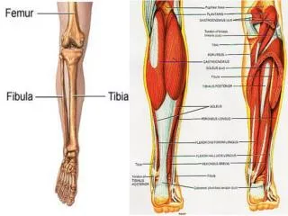
The femur The bone that holds it all together • Anterior landmarks • Head • Neck • Greater trochanter • Lesser trochanter • Lateral condyle • Medial condyle • Intercondylarfossa • Medial epicondyle • Lateral epicondyle