Light
O 2. CO 2. Light. Sugar. H 2 O. O 2. H 2 O and minerals. CO 2. 32. 24. 42. 40. 29. 16. 19. 11. 21. 27. 3. 8. 34. 6. 14. 13. Shoot apical meristem. 26. 1. 22. 5. 9. 18. Buds. 4. 10. 2. 17. 31. 12. 7. 23. 15. 25. 20. 28. 1 mm. Cell wall.

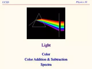
Light
E N D
Presentation Transcript
O2 CO2 Light Sugar H2O O2 H2O and minerals CO2
32 24 42 40 29 16 19 11 21 27 3 8 34 6 14 13 Shoot apical meristem 26 1 22 5 9 18 Buds 4 10 2 17 31 12 7 23 15 25 20 28 1 mm
Cell wall Apoplastic route Cytosol Symplastic route Transmembrane route Key Plasmodesma Apoplast Plasma membrane Symplast
EXTRACELLULAR FLUID CYTOPLASM H+ H+ S H+ H+ H+ Hydrogen ion H+ H+ H+ S S H+ H+ H+ H+ H+ H+ H+ S S S H+ H+ H+ Proton pump H+ Sucrose (neutral solute) H+/sucrose cotransporter (a) H+ and membrane potential (b) H+ and cotransport of neutral solutes H+ H+ NO3− NO3− H+ K+ Potassium ion H+ H+ K+ H+ Nitrate K+ H+ K+ H+ NO3− K+ NO3− NO3− NO3− K+ K+ H+ H+/NO3− cotransporter H+ H+ Ion channel (d) Ion channels (c) H+ and cotransport of ions
−0.7 MPa P S 0 0 P S −0.9 0 0 MPa Initial flaccid cell: P S −0.7 0 0.4 M sucrose solution: Pure water: Turgid cell at osmotic equilibrium with its surroundings Plasmolyzed cell at osmotic equilibrium with its surroundings P S P S −0.9 0 0.7 −0.7 −0.9 MPa −0.9 MPa 0 MPa (b) Initial conditions: cellular environmental (a) Initial conditions: cellular environmental
Wilted Turgid
Technique Control: Solution containing all minerals Experimental: Solution without potassium
Healthy Phosphate-deficient Potassium-deficient Nitrogen-deficient
Soil particle K+ K+ Ca2+ Mg2+ Ca2+ K+ H+ H+ H2CO3 H2O + CO2 HCO3−+ Root hair Cell wall
ATMOSPHERE N2 SOIL N2 N2 ATMOSPHERE Nitrate and nitrogenous organic compounds exported in xylem to shoot system Proteins from humus (dead organic material) SOIL Nitrogen-fixing bacteria Microbial decomposition Amino acids Denitrifying bacteria NH3 (ammonia) Ammonifying bacteria NH4+ H+ (from soil) NO3− (nitrate) NO2− (nitrite) NH4+ (ammonium) Nitrifying bacteria Nitrifying bacteria Root
Nodules Roots
Cortex Mantle (fungal sheath) Epidermis Epidermal cell Endodermis (Colorized SEM) Fungal hyphae between cortical cells 1.5 mm Mantle (fungal sheath) (LM) 50 m (a) Ectomycorrhizae Cortex Epidermis Cortical cell Endodermis Fungal vesicle Fungal hyphae Casparian strip 10 m Root hair Arbuscules Plasma membrane (LM) (b) Arbuscular mycorrhizae (endomycorrhizae)
Results Experiment 300 200 Increase in plant biomass (%) 100 0 Uninvaded Invaded Sterilized uninvaded Sterilized invaded Soil type 40 30 Mycorrhizal colonization (%) 20 Seedlings 10 Sugar maple 0 Red maple Uninvaded Invaded White ash Soil type
Parasitic plants Dodder, a nonphoto- synthetic parasite (orange) Indian pipe, a nonphoto- synthetic parasite of mycorrhizae Mistletoe, a photosynthetic parasite
Carnivorous plants Sundew Pitcher plants Venus flytraps
4 5 4 3 3 5 4 2 2 1 1 5 Casparian strip Endodermal cell Pathway along apoplast Pathway through symplast Apoplastic route Casparian strip Plasma membrane Apoplastic route Symplastic route Vessels (xylem) Root hair Symplastic route Transmembrane route Epidermis Endodermis Vascular cylinder (stele) Cortex The endodermis: controlled entry to the vascular cylinder (stele) Transport in the xylem
Xylem Cuticle Upper epidermis Microfibrils in cell wall of mesophyll cell Mesophyll Air space Lower epidermis Cuticle Stoma Microfibril (cross section) Water film Air-water interface
−0.8 MPa −0.6 MPa −0.3 MPa Xylem sap Outside air Mesophyll cells −100.0 MPa = Stoma Leaf (air spaces) Water molecule = −7.0 MPa Atmosphere Transpiration Leaf (cell walls) Adhesion by hydrogen bonding −1.0 MPa = Xylem cells Cell wall Water potential gradient Trunk xylem Cohesion by hydrogen bonding Cohesion and adhesion in the xylem Water molecule Root hair Trunk xylem Soil particle Water Soil Water uptake from soil
Guard cells flaccid/Stoma closed Guard cells turgid/Stoma open Radially oriented cellulose microfibrils Cell wall Vacuole Guard cell (a) Changes in guard cell shape and stomatal opening and closing (surface view) H2O H2O H2O H2O H2O K+ H2O H2O H2O H2O H2O (b) Role of potassium ions (K+) in stomatal opening and closing
Oleander (Nerium oleander) Ocotillo (Fouquieria splendens) Upper epidermal tissue Thick cuticle 100 m Lower epidermal tissue Trichomes (“hairs”) Stoma Crypt Old man cactus (Cephalocereus senilis)
Apoplast Symplast Companion (transfer) cell High H+ concentration Mesophyll cell Cotransporter H+ Cell walls (apoplast) Proton pump Sieve-tube element Plasma membrane S Plasmodesmata Sucrose H+ H+ S Phloem parenchyma cell Bundle- sheath cell Mesophyll cell Low H+ concentration (b) A chemiosmotic mechanism is responsible for the active transport of sucrose. (a) Sucrose manufactured in mesophyll cells can travel via the symplast (blue arrows) to sieve-tube elements.
1 1 2 2 3 4 3 4 Sieve tube (phloem) Source cell (leaf) Vessel (xylem) Loading of sugar H2O Sucrose H2O Uptake of water Bulk flow by negative pressure Bulk flow by positive pressure Unloading of sugar Sink cell (storage root) Recycling of water Sucrose H2O