Download
1 / 56
560 likes | 704 Views
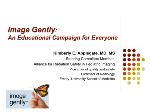
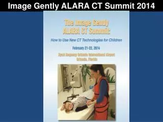
IMAGE GENTLY AND NEW JERSEY CT DOSE PROJECT UPDATE. New Jersey Medical Physicist Meeting March 6, 2008. Image Gently Campaign. Launched by The Alliance for Radiation Safety in Pediatric Imaging on January 22, 2008 Formed in 2007 Made up of 13 Healthcare Organizations.

E N D
IMAGE GENTLY ANDNEW JERSEY CT DOSE PROJECT UPDATE New Jersey Medical Physicist Meeting March 6, 2008
Image GentlyCampaign Launched by The Alliance for Radiation Safety in Pediatric Imaging on January 22, 2008 Formed in 2007 Made up of 13 Healthcare Organizations
Who is the Alliance? • American Association of Physicists in Medicine* • American College of Radiology* • American Society of Radiologic Technologists * • The Society for Pediatric Radiology* • American Academy of Pediatrics • American Osteopathic College of Radiology • American Registry of Radiologic Technologists • American Roentgen Ray Society • Association of University Radiologists • Conference of Radiation Control Program Directors • National Council on Radiation Protection • Radiological Society of North America • Society of Computed Body Tomography and Magnetic Resonance * Founding member
What is Image Gently Goal? Goal:“Is to change the practice” • To raise awareness of the opportunities to lower radiation dose in the imaging of children Focus: Pediatric CT Procedures Key Points: • One Size Does Not Fit All … • Child Size mAs and kVp • Radiation Matters! What we do now, lasts their lifetime • More is not better • When we Image, let’s Image Gently
How Can Practice be Changed? Answer: EDUCATION Web (www.imagegently.org) contains educational sites for: • Parents • Physicians • Medical Physicists • Technologists • The Press
Parents Be your Child’s Advocate Ask Questions regarding: • The use of alternative imaging modalities • Benefit and Risk and Ways to Reduce Dose Ask Questions about the CT Facility: • Is the facility ACR Accredited? • Are technologists credentialed? • Are interpreting physicians board certified radiologists or pediatric radiologists?
Physicians, Physicists and TechnologistsHow to Change your Practice: Increasing your awareness for the need to decrease radiation dose to Children. Be committed to make change and work as a team with parents, physicians, physicists and technologists to decrease dose. Take the web Pledge.
Know your practice standards • Use ACR’s Appropriateness Criteria for ordering procedures. • Establish CT Protocols for both adult and pediatric procedures. • For Pediatric Protocols: • Child-size mAs and kVp • Limit scan to only the indicated area • A single scan is usually adequate (Pre, post and delayed contrast scans ( i.e., contrast-enhanced multi-phase scanning) rarely add additional information.)
Pediatric CT Protocol Procedure and Excel Worksheet SUMMARY OF INSTRUCTIONS TO ESTABLISH SUGGESTED PROTOCOLS Establish Adult Protocols for Abdomen and Head where the CTDI(vol) are below ACR’s reference levels of 25 mGy for Abdomen and 75 mGy for Head. There are 2 worksheets: • Pediatric Abdomen and Thorax • Pediatric Head
The Abdomen - Thorax worksheet has: • 5 pediatric ages and “Average” PA Thicknesses and 3 adult size “Average” Thicknesses • The “Med Adult” = 25 cm is the Adult Protocol’s mAs • For all other a “mAs Reduction Factor” (RF) is listed. • The RF for 5yrs of age (14 cm) is 0.59 The Head worksheet has: • 3 pediatric ages and “Average” PA Thicknesses and 1 Med Adult size. Enter the mAs used for the Adult protocol in the Med Adult as the Baseline . Using the Baseline mAs and the “mAs Reduction Factors”, the estimated mAs for each size is calculated.
Worksheet/Procedure Limitations Worksheet may not work for all CT types Worksheet cannot be used if kVp is changed • In 2006, the AAPM was discussing the issue of decreasing kVp from 120 to 80 for pediatric CT procedures to improve image contrast. AAPM Report #96 supports a lower kVp but no suggestions were made. Procedure does not discuss the importance of Pitch on dose reduction.
Limitations Greater emphasis should have been placed on the use of mA Modulation, if equipped. AAPM Report #96 reports that the typical reduction in mAs from the adult abdomen to an infant is 4 to 5 times (Image Gently’s reduction factor is 0.43 to 0.51).
Why is Monitoring CT DoseImportant? NCRP SC-6-2 Medical Subgroup reported its preliminary results in April 2007: • 67 million CT procedures are performed in the United States annually. 7 million are pediatric. 33% of which are performed on children under the age of 10. • In 1993, 18.3 million CT procedures were performed. The annual growth of CT procedures increased >10%/yr, whereas the U.S. population increased by < 1%/yr. • 73% of CT abdomen and pelvis procedures are performed on patients between the ages of 45 and 84 which account for only 34% of the U.S. population. • CT procedures account for 16% of all medical rad/fluoro procedures performed, but contribute 63% of the dose delivered to patients.
Studies conducted by Duke University and The University of North Carolina at Chapel Hill on the use of CT in ERs reveal significant increase use of CT: • From 2000 to 2005 in adult patients. (Some stats: Head: 51%, C-Spine: 463%, Chest: 226%, Abdomen: 72%) and • From 2000 to 2006 in pediatric patients (Some stats: Head: 66%, C-Spine: 731%, Chest: 675%, Abdomen: 104%) • In the 11th Edition of the “Report of Carcinogens” (2005) published by the USDHHS, x-ray and gamma are listed as “Known Human Carcinogens” (first –time recognized) • According to a study published in JAMA Vol. 298 No.3 on July 18, 2007, the estimated lifetime cancer risk from a CT Coronary Angiography (CTCA) is between 1 in 143 to 1 in 3261 depending on the patient’s age and gender.
According to the BEIR VII Report, the estimated lifetime cancer (i.e., solid and leukemia) risk from a 100 mSv dose is approximately 1 individual in 100 persons. • According to the ACR, “Many CT scans have an effective dose estimates in the range of 15-25 mSv for a single study.”
Typical CT & Non-CT Effective Doses (Source: AAPM Report #96)
NJ CT DOSE PROJECT History and Second Data Set
New Jersey CT Dose History • In 2003-2004, a mail in survey conducted by the Bureau of NJ CT facilities revealed a need to standardize the method of calculating and reporting CT doses. • Since measuring CT dose is a required test to be performed by medical physicists as part of NJ’s Annual CT QC survey, in November 2004, Bureau met with NJ medical physicists regarding this issue.
2004 MeetingOutcomesPhysicists Agreed with the BRH • A standardized method for calculating and reporting CT dose should be used. • ACR’s CT Accreditation Program’s method and set up procedures for calculating CT dose should be used. • BRH to develop a form to report CT dose. • CT dose should be calculated and reported for Adult Head, Adult Abdomen and Pediatric Abdomen. • Patient scan protocols should be used.
2004 Meeting Outcomes • NJ should apply ACR’s CTDI(w) reference levels as its reference levels for CTDI(vol). • This will account for pitch from helical scanning
Current NJ Reference Levels Same Reference Levels established by the ACR BUT NJ uses CTDI (vol) and not CTDI(w)
March 2006 New Jersey Medical Physicists Meeting • The Bureau met with medical physicists to review CT doses collected from December 2004 to February 2006. • This Data Set included 396 CT doses were collected on 141 CT scanners. (About 40% of all registered CT scanners) • The following Baseline was established:
Baseline CTDI(vol) Doses (mGy)Data is Established The 80th percentile is recommended by the AAPM (May 2005)
2006 Meeting Outcomes • For all three procedures, NJ’s Baseline mean CTDI(vol) doses were below ACR’s CTDI(w) reference levels. • Facilities experiencing difficulties staying below ACR’s reference level for Adult Head. • New Jersey’s CT Dose Report updated to provide better statistical analysis. • Education is needed in Multi-Slice CT and pediatric abdomen protocols.
Analysis of Data Set 2 CT Dose Data • Demographic Information: • Data Set 2 includes CT doses collected from March 2006 to August 2007. • A total of 334 CT Doses were collected on 115 CT scanners. (About 34% of all registered CT scanners) • Compared to the Baseline, this Data Set consists of 62 fewer doses on 26 fewer units.
Comparison of Slice Type Data Baseline Data Set 2
Comparison of Adult Head Mean CTDI(vol) Doses Only 1 dose collected for 6 and 32 Slice Units
Comparison of Adult Abdomen Mean CTDI(vol) Doses Only 1 dose collected for 6 and 32 Slice Units
Comparison of Pediatric Abdomen Mean CTDI(vol) Doses Only 1 dose collected for 6 and 32 Slice Units
Comparison of CT Doses Above NJ’s CTDI(vol) Reference Levels(in %)
Analysis of the 19% (22) High Adult Head Doses by Slice Type
Analysis of the 3.5% (4) High Adult Abdomen Doses by Slice Type
Analysis of the 2% (2) High Pediatric Abdomen Doses by Slice Type
Analysis of All (28) High CTDI(vol) by Slice Type for All Procedures
NJ Mean mAs Reduction From Adult Abdomen Image Gently suggests a reduction factor of 0.59 from the average (25 cm) adult to a 5 yrs old (14 cm) child. Note: Comparison cannot be made if kVp is different. For all slice types, the actual Pediatric mAs used was lower than the Adult . When kVp was the same, the actual mAs used for pediatric procedures were either equal to or lower than the calculated estimated mAs. (See Slice #s 6, 8, 32 and 64). The lower kVp in 2 slice CT units, resulted in a higher actual mAs used than the calculated estimated mAs.
ACR’s New Dose RLs and Limits • Effective January 1, 2008 • CTDI(vol) will be used to determine Dose compliance. • Establishes Dose Reference Levels and Pass/Fail Limits. • Major Dose Limit Changes: • Adult Head increased from 60 mGy to 80 mGy • Adult Abdomen decreased from 35 mGy to 30 mGy • Using Data Set 2, these new values will have the following effects:
Data Set 2: Adult Head Doses Above ACR’s Values Note: Old is prior to 1/1/08 Based on 115 collected doses
Data Set 2: Adult Abdomen Doses Above ACR’s Values Note: Old is prior to 1/1/08 Basedon 115 collected doses
Data Set 2: Pediatric Abdomen Doses Above ACR’s Values Note: Old is prior to 1/1/08 Based on 104 collected doses
Data Set 2 Observations • 16 and greater slice scanners may make up the majority of registered scanners in NJ. • As compared to the Baseline, the mean CTDI (vol) dose decreased in adult and ped. abdomen and the 80th %tile doses decreased for all procedures. Most significant decrease was in Pediatric Abdomen which decreased by 20%. • The mean dose increase in Adult Head may be contributed to the anticipated increase in ACR’s limit to 80 mGy.