Resonant waveguide grating biosensor for living cell sensing
180 likes | 394 Views
Resonant waveguide grating biosensor for living cell sensing. Ye Fang, Ann M. Ferrie, Norman H. Fontaine, John Mauro, and Jitendra Balakrishnan Biochemical Technologies, Science and Technology Division, Corning Incorporated Biophysical Journal, June 2006. Presentation Outline. Motivation

Resonant waveguide grating biosensor for living cell sensing
E N D
Presentation Transcript
Resonant waveguide grating biosensor for living cell sensing Ye Fang, Ann M. Ferrie, Norman H. Fontaine, John Mauro, and Jitendra Balakrishnan Biochemical Technologies, Science and Technology Division, Corning Incorporated Biophysical Journal, June 2006 Jeremy Colson, Boston University
Presentation Outline • Motivation • Background: assays, RWGs • Methods • Vertical and horizontal mass distributions • Results • Cell adhesion and spreading • Cell detachment • Conclusion • Where are they now? Jeremy Colson, Boston University
Assays • Procedure to determine the concentration of a component part of a mixture • Cell-based • More complex, less specific • Useful for functional information • Pathway activation • Toxicity • Phenotypic responses • Need for label-free Jeremy Colson, Boston University
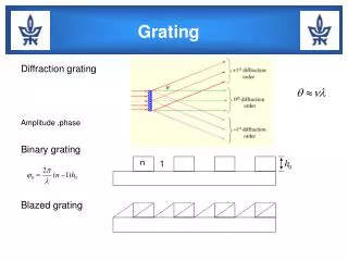
Resonant Waveguide Gratings & MRCAT Jeremy Colson, Boston University
Mass redistribution detection method • Intensity as a function on incident angle • Resonant peaks • Vertical mass redistribution • Shift in resonant peak • Lateral mass redistribution • Changes in peak shape (PWHM) Jeremy Colson, Boston University
Effective refractive index 1 response unit = 5.82x10⁻⁴ Jeremy Colson, Boston University
Vertical mass redistribution Specific refractive index increment α = .0018 per 100ml/g (protein) .0016 per 100ml/g (other, Na) Jeremy Colson, Boston University
Comments on ΔN • Primarily sensitive to vertical mass redistribution (DMR) • Directly a function of changes in protein concentration due to protein relocation (rather than ion mobilization) mediated by a stimulation • Relocation of a target or complex of certain mass near the sensor surface contributes more to the overall response than those further away • Optical signature is an integrated signal that is a sum of contributions from mass redistribution occurring at different distances away from sensor surface Jeremy Colson, Boston University
Horizontal mass movement • Lateral inhomogeneity does not affect refractive index • Lateral inhomogeneity does affect shape of resonant peaks Jeremy Colson, Boston University
Cell adhesion and spreading • Human epidermoid carcinoma cells (A431) in 5% FBS • At room temp adhesion not optimal • Cells interact with surface through multiple steps • Spreading step increases the mass w/in sensing volume Jeremy Colson, Boston University
Cell spreading inhibitor added • 100nM vincristine • Reduced kinetics of cell spreading • Initial steps primarily affected • Are the effects of vincristine limited to first 14 hours? Jeremy Colson, Boston University
Cell detachment • Trypsin – pancreatic serine protease with substrate specificity based on positively charged lysine and argenine side chains • Used for cell detachment • A431 cells, 95% confluency Jeremy Colson, Boston University
Low-doses: cell signaling • Presence of P-DMR: cell signaling • Slight N-DMR: insignificant cell detachment • => activation of endogenous protease-activated receptors that lead to typical Gq signaling Jeremy Colson, Boston University
Conclusions • Optical signatures are integrated responses that can be used to examine cells in native environments label-free • Systematic investigation of cell processes • Adhesion • Detachment • Cell signaling: EGFR and Bradykinin B2 receptors Jeremy Colson, Boston University
Back to Epic… Jeremy Colson, Boston University
Thank you! Jeremy Colson, Boston University