Download
1 / 14
140 likes | 245 Views
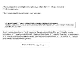
The major question resulting from these findings is how these two subsets of memory T cells are generated. Three models of differentiation have been proposed:. Two subsets of memory T lymphocytes with distinct homing potentials and effector functions

E N D
The major question resulting from these findings is how these two subsets of memory T cells are generated. Three models of differentiation have been proposed: Two subsets of memory T lymphocytes with distinct homing potentials and effector functions FEDERICA SALLUSTO*, DANIELLE LENIG*, REINHOLD FÖRSTER†, MARTIN LIPP† & ANTONIO LANZAVECCHIA* Nature401, 708 - 712 (1999) In vitro stimulation of naive T cells resulted in the generation of both TCM and TEM cells, whereas stimulation of TCM cells resulted in their efficient differentiation to TEM cells. These data were consistent with a linear differentiation model in which naive T cells differentiate first to TCM and then to TEM cells, which were considered end-stage cells. Naive effectorsTCM TEM
+ + Migratory properties of naive, effector, and memory CD8(+) T cells Weninger W, Crowley MA, Manjunath N, von Andrian UH. J. Exp. Med. 194, 953-966 (2001) According to Manjunath et al., the duration of antigenic stimulation and the type and amount of cytokines present during priming lead either to fully differentiated effector cells that home to peripheral tissues (blue) or to cells that are devoid of effector function and home to lymph nodes (green). In the system used by Manjunath et al., these two cell types can be identified according to the differential expression of the T-GFP marker transgene and the lymph node–homing receptor CCR7. Both cell types are maintained in the memory pool (dotted arrows) and, upon secondary challenge, mediate immediate protection in nonlymphoid tissues or secondary responses in lymph nodes. The repertoires of circulating human CD8+ central and effector memory T cell subsets are largely distinct. Baron V., Bouneaud C., Cumano A., LimA., Arstila T.P., KourilskyP., FerradiniL.,and Pannetier C. Immunity 18, 193-204 (2003) Baron et al. analyzed the composition and dynamics of the CD8+ T cell repertoire of these subsets within the peripheral blood of four healthy individuals. Both subsets had largely distinct and autonomous TCRV repertoires. Their composition remained stable over a 9 month period, during which no cell passage between these subsets was detected despite important size variation of several clones. In one donor, four out of six TCRV clonotypes specific for the influenza A virus were detected in the central subset only, while the two others were shared. Altogether, these observations suggest that most effector memory T cells may not have derived from the central memory subset.
Lineage relationship and protective immunity of memory CD8 T cell subsets Wherry, J.E. et all. Nature Immunol 4, 225-234 (2003) • We will examine in details a recent publication which addresses the following points: • The lineage relationships between TCM and TEM • Which memory T cell subsets has the greater capacity to persist long-term in vivo and undergo • homeostatic proliferation • Because TEM are located in non-lymphoid tissues, it has been proposed that they may • represent a more effective population for protection from reinfection. They will directly compare • in vivo the protective capacity of the two subsets of memory T cells. Their tools: Tetramers that allow the detection of T cells specific for a peptide of gp30/LCMV presented by Db. For some experiments they will also use TCR transgenic T cells that are specific for the same peptide of gp30/LCMV presented by Db. These TCR transgenic T cells will be transferred into normal mice.
Normal behavior of gp-33 specific T cells upon infection Effector cells are highly cytotoxic directly ex vivo The lytic capacity decreases gradually over time Figure 1.Characterization of effector and memory T cells. (a) Viral load and Db-gp33–specific CD8 T cell numbers in the spleen after LCMV Armstrong infection of B6 mice. (b) Cytotoxicity of Db-gp33–specific CD8 T cells at days 8, 15 and 30 post LCMV Armstrong infection. E:T was 2:1 in all cases. (c) Intracellular granzyme B staining of Db-gp33–specific CD8 T cells at 8, 15 and 125 (memory) d.p.i. The open histogram indicates naive cells. (d) In vivo homing of naive, effector and memory T cells. Naive, effector (8 d.p.i.) and memory (60 d.p.i.) P14 cells (Thy1.1+) were adoptively transferred into naive B6 (Thy1.2+) recipients. After 12 h the number of donor (Thy1.1+) gp33-specific CD8 T cells was determined in the indicated organs by flow cytometry. 8 d.p.i 60 d.p.i Correlates with decreased expression of GrB Homing properties Naïve: lymphoid organs not non-lymphoid organs Effector: reduced ability to home to LN, gain homing to non-lymphoid organs Memory: in contrast to naïve home to non-lymphoid organs In contrast to effectors regain ability to enter LN
Figure 2. Characterization of memory T cell subsets. (a) LCMV Db-gp33–specific memory CD8 T cells (2–3 months p.i.) were costained for CD62L expression (histogram is gated on CD8+Db-gp33+ cells). (b) Db-gp33–specific memory T cells (2–4 months p.i.) were costained for CD62L and CCR7 expression (left) or CD62L and CD27 expression (right). Plots are gated on CD8+Db-gp33+ cells. (c) Phenotypic analysis of CD62Lhi and CD62Llo subsets of Db-gp33–specific memory CD8 T cells. Histograms are gated on either CD62Lhi (top) or CD62Llo (bottom) CD8+Db-gp33+ memory cells (1–2 months p.i.). Open histograms indicate naive cells. Memory gp-33 specific CD8+ T cells form TCM and TEM
In vitro migration of TCM to chemokines correlates with capacity to home to LN TCM in LN TEM in non-lymphoid tissues Figure 2. Characterization of memory T cell subsets. (d) LCMV gp33-specific memory (60 d.p.i.) cells from LN, liver and lung of P14 chimeras were stained for CD62L expression. Histograms are gated CD8+Db-gp33+ memory cells. Similar results were observed for normal B6 mice. (e) Splenocytes from P14 LCMV-immune chimeras (> 30 d.p.i.) were added to a transwell plate and migration in the presence or absence of added chemokine (100 nM) was assessed. (f) IFN-, TNF- and IL-2 production by gp33-specific TCM and TEM P14 splenocytes separated using magnetic beads (92% and 97% pure, respectively) was assessed by ICS following gp33 peptide stimulation. (g) ICS by TCM from LN and TEM from the liver. (h) A 5- and 18-h gp33-specific 51Cr release assay using splenic TCM and TEM purified as in (f) (left) or using memory T cells from spleen versus liver. All immune mice used in functional experiments were > 30 d.p.i. Background lysis in the absence of gp33 peptide was similar for TCM and TEM and has been subtracted. (i) Granzyme B staining of memory T cell subsets from the spleen. TCM are gated on CD62Lhi and TEM on CD62Llo Db-gp33+CD8 T cells at 60 d.p.i. Open histograms indicate staining with an isotype control antibody. TCM and TEM both acquire effector functions upon restim. In vitro (in contrast to previous reports).
Figure 3.Protective immunity by memory T cell subsets. (a) Thy1.1+ gp33-specific P14 transgenic memory cells were generated by infecting B6 chimeric mice containing P14 cells with LCMV (Armstrong) or recombinant LM expressing the gp33 epitope (LMgp33). CD62Lhi or CD62Llo splenocytes 1- to 2-months p.i. were purified by flow cytometry or magnetic bead separation. Equal numbers of CD62Lhi or CD62Llo gp33-specific P14 cells were adoptively transferred to separate naive mice. Two days later, recipients were challenged as indicated. n = 3–6 mice in all groups for all experiments. (b) Purity of CD62Lhi and CD62Llo populations before transfer. (c) To determine the number of TCM and TEM present in recipient mice following adoptive transfer, the number of Db-gp33+Thy1.1+CD8+ cells in the indicated organs was measured by flow cytometry (2 d after transfer into naive Thy1.2+ mice). (d) Control of LCMV clone-13 infection by TCM or TEM (7.5 104 of each) following intravenous (i.v.) challenge. (e) Control of VVgp33 infection by TCM or TEM (2.5 105 of each) following i.p. challenge. VV ovary titers were determined on day 5 (TEMversus TCM, P = 0.08). Given the similarities in effector function, they will determine whether TCM and TEM differ in their ability to confer protective immunity
Figure 3.Protective immunity by memory T cell subsets. (f) Control of LCMV clone-13 infection by LMgp33-induced TCM or TEM (1 105 of each) following i.v. challenge. Day 8 serum viral titers are shown (TEMversus TCM, P = 0.02). (g) Induction of DTH response by LMgp33-induced TCM or TEM (2 105 of each) following footpad injection of LCMV clone-13. Footpad thickness was measured daily. (h) Control of VVgp33 infection by LCMV-induced TCM, TEM or lung-derived TEM (3 105 of each) following i.n. challenge (TEM spleen versus TCM, P = 0.04; TEM lung versus TCM, P = 0.003). VV lung titers were determined on day 5. For all protection experiments at least two doses of cells were transferred. The number of memory cells transferred in the experiments shown is indicated in parentheses above. On a per cell basis, TCM more effectively control viral replication than TEM . Independent of the route of infection or the site of infection, (in contrast to expectations)
respiratory challenge systemic challenge Figure 4.Antigen-driven proliferation of memory T cell subsets. (a) In vivo T cell expansion following systemic challenge. Db-gp33+CD8+ T cells were enumerated in spleen, PBMC, LN and liver of TCM and TEM recipients 5 d after i.p. VVgp33 challenge (Fig. 3e). All Db-gp33+CD8+ T cells were donor derived (Thy1.1+; data not shown). TCM recipients had significantly more total Thy1.1+Db-gp33+CD8+ T cells in all locations examined (P < 0.05). (b) In vivo T cell expansion following respiratory challenge. Thy1.1+ (donor) Db-gp33+CD8+ T cells were enumerated in spleen, PBMC, LN and lung of TCM and TEM recipients 5 d after i.n. VVgp33 challenge (Fig. 3h). TCM recipients had significantly more total Thy1.1+Db-gp33+CD8+ T cells in lung, spleen and PBMC (P < 0.05). TCM show greater expansion in vivo, can be explained by the fact that T cell priming occurs in LN and TCM in contrast to TEM localize to the LN.
TCM have a greater intrinsinc advantage over Tem to proliferate following antigenic stimulation. IL-2? The increased capacity of TCM to provide protection may be the result of generating a larger number of secondary effectors. TCM convert to TEM following antigen challenge Figure 4.Antigen-driven proliferation of memory T cell subsets. (c) In vitro proliferation of TCM and TEM P14 cells in response to gp33 peptide. The mean division number for TCM and TEM was 3.4 and 2.0, respectively. No division was observed in the absence of gp33 peptide (data not shown). (d) Five days after i.p. VVgp33 challenge, recipients of TCM or TEM were sacrificed and the expression of CD62L on secondary effectors in the spleen, PBMC, LN and liver was assessed by flow cytometry. The phenotype of the pretransfer populations of gp33-specific memory cells are shown at the top. Data are representative of 3–4 mice/group.
TCM, TEM: death or conversion? TEM convert to TCM after transfer in vivo into naïve recipients Figure 5.Lineage relationship between memory T cell subsets. (a) The number of total and CD62Lhi and CD62Llo memory Db-gp33+CD8+ T cells and the percentage of CCR7+ and CCR7- Db-gp33+ CD8 T cells in the spleens of LCMV immune P14 chimeric mice are plotted over time. n = 2–4 mice/time point. (b) Column-purified CD62Lhi or CD62Llo Db-gp33+ memory T cells were adoptively transferred into separate naive mice. After 25 d, CD62L expression on splenic Db-gp33+CD8+ T cells of recipients was determined.
Higher homeostatic proliferation of TCM TEM convert to TCM after transfer in vivo into naïve recipients Long term persistence of memory T cells is in the form of TCM. TEM do not seem to be a permanent population, but rather convert to TCM and in so doing acquire the ability to undergo efficient, antigen-independent homeostatic proliferation Figure 5.Lineage relationship between memory T cell subsets. (c) Purified TCM and TEM cells were CFSE-labeled and transferred into separate naive recipients (nonirradiated). Division of the transferred Thy1.1+ P14 memory cells was assessed after 30 d. (d) Purified TCM or TEM Db-gp33+ cells were CFSE labeled and transferred to naive mice (nonirradiated). After 1 and 30 d, CD62L expression was examined as a function of division. Dot plots are gated on Thy1.1+ P14 memory CD8 T cells from the spleen. (e) CCR7 and CD27 expression was examined as a function of cell division on transferred TEM cells 30 d post transfer. (f) Purified TCM and TEM Db-gp33+ CFSE-labeled memory CD8 T cells were transferred separately into naive irradiated recipients. Division of the transferred Thy1.1+ P14 memory cells was analyzed after 8 d.
Figure 5.Lineage relationship between memory T cell subsets.(g) LCMV immune (85 d.p.i.) mice were fed BrdU in their drinking water for 8 d and splenocytes were stained for BrdU incorporation. Db-gp33 tetramer staining versus CD62L is shown for gated CD8 cells. Histograms are gated on Db-gp33+CD8+CD62Lhi (top) or CD62Llo (bottom) memory T cells. (h) Db-gp33+CD8+ memory T cells (30 d.p.i.) from a P14 chimera were stained for CD62L expression and the percentage of cells increased in size was indicated by high forward scatter was assessed. Plots are gated on Db-gp33+CD8+ cells. (i) IL-2 production by Db-gp33–specific memory CD8 T cells was assessed at the indicated times p.i. by intracellular cytokine staining following gp33 peptide stimulation. Naive effectors TEM TCM Ag TEM
Figure 6.The effect of HD versus LD infection on the duration of TEM to TCM conversion. (a) P14 chimeras were infected with a LD (500 c.f.u.) or HD (3 104 c.f.u.) of LMgp33 or with LCMV and the percentage of gp33-specific CD8 T cells (P14 cells) that were CD62Lhi or CD62Llo in the blood was determined longitudinally in individual mice. (b) Naive Thy1.1+ P14 chimeras were infected with LD LMgp33 and separate naive Thy1.2+ P14 chimeras were infected with LCMV. After 8 d.p.i. spleens were harvested, CD8 T cells column purified (both > 96% pure) and CD8 T cell populations from LMgp33 and LCMV infected mice mixed and transferred into the same recipients. Reexpression of CD62L was monitored on LD LMgp33-induced (Thy1.1+) and HD LCMV-induced (Thy1.2+) P14 cells parked in the same mice. P14 cells in the PBMC were analyzed over time. Data are representative of four independent experiments. Does the differentiation of TEM to TCM is affected by the magnitude of the infection? The conversion occurs more rapidly in LD than HD. Is it programmed during the initial phase of priming or the result of the persisting Ag?