EVAR Endofuite de Type II Embolisation directe
200 likes | 399 Views
EVAR Endofuite de Type II Embolisation directe. Jean-Paul Beregi, Serge Ovtchinnikoff, Cornelia Freitag, Romain Bechet, Francesco Macri Radiologie et Imagerie Médicale CHU de Nîmes, 30029 Nimes Cedex 9 jean.paul.beregi@chu-nimes.fr. S Haulon and JP Beregi.

EVAR Endofuite de Type II Embolisation directe
E N D
Presentation Transcript
EVAR Endofuite de Type IIEmbolisation directe Jean-Paul Beregi, Serge Ovtchinnikoff, Cornelia Freitag, Romain Bechet, Francesco Macri Radiologie et Imagerie Médicale CHU de Nîmes, 30029 Nimes Cedex 9 jean.paul.beregi@chu-nimes.fr
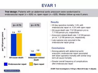
S Haulon and JP Beregi • Embolization of type II endoleaks after aortic stent-graft implantation: technique and immediate results. Haulon S, Tyazi A, Willoteaux S, Koussa M, Lions C, Beregi JP. J Vasc Surg. 2001 Oct;34(4):600-5. • Prospective evaluation of magnetic resonance imaging after endovascular treatment of infrarenal aortic aneurysms. Haulon S, Lions C, McFadden EP, Koussa M, Gaxotte V, Halna P, Beregi JP. Eur J Vasc Endovasc Surg. 2001 Jul;22(1):62-9. • Diagnosis and treatment of type II endoleak after stent placement for exclusion of an abdominal aortic aneurysm. Haulon S, Willoteaux S, Koussa M, Gaxotte V, Beregi JP, Warembourg H. Ann Vasc Surg. 2001 Mar;15(2):148-54. Epub 2001 Mar 1. jean.paul.beregi@chu-nimes.fr
Technique (au début) • Anesthésie Générale • Décubitus ventral • Scanner • Aiguille 22G + micro-KT jean.paul.beregi@chu-nimes.fr
Cas Clinique jean.paul.beregi@chu-nimes.fr
Expérience personnelle • Lille / Nîmes • N=15 cas • Anesthésie locale +/- sédation en Externe • Aiguille métallique • Micro-KT • Micro-guide • Sous Scanner • Décubitus / Procubitus / Latéral • 1 à 3 ponctions • Onyx : 1 à 3 flacons par séance • Nécessité de savoir répéter l’embolisation jean.paul.beregi@chu-nimes.fr
Quel agent d’embolisation ? • Coils • Hysto-acryl • Onyx : Ethylene-Vinyl-Alcohol Copolymer jean.paul.beregi@chu-nimes.fr
Difficulté approche translombaire • Analyse scanographique • Structure adjacente • Ponction directe angulée ? • Repérage • Module scanographique interventionnel • Association avec amplificateur jean.paul.beregi@chu-nimes.fr
Suivi • Scanner en général (ou ED) • Si fuite persistante avec diamètre stable ou qui augmente, savoir répéter l’embolisation jean.paul.beregi@chu-nimes.fr
Littérature jean.paul.beregi@chu-nimes.fr
Conclusion • Une endofuite aortique doit être abordée comme une MAV ! • Embolisation • Nidus : anévrisme • Artère afférente • Artères efférentes • Voie TL > voie TF • Echec TF • Matériel adéquat et logistique spécifique jean.paul.beregi@chu-nimes.fr