Download
1 / 40
530 likes | 821 Views
INFRATEMPORAL FOSSA Contents Mandibular nerve Otic ganglion. M. ASIF BMLT., M.Sc(medical anatomy) Dept Of Anatomy. INFRATEMPORAL FOSSA.

E N D
INFRATEMPORAL FOSSA Contents Mandibular nerve Otic ganglion M. ASIF BMLT., M.Sc(medical anatomy) Dept Of Anatomy
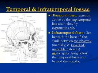
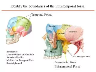
INFRATEMPORAL FOSSA • Region situated below the middle cranial fossa of the skull, intervenes between the pharynx and the ramus of the mandible
BOUNDARIES • Anterior- maxilla • Posterior- styloid process • Medial- lateral pterygoid plate • Lateral- ramus & coronoid process • Above- Infratemporal surface of greater wing of sphenoid • Below- open
COMMUNICATIONS • Infront- orbit through inferior orbital fissure • Medial- Pterygopalatine fossa through pterygopalatine fissure • Above & lateral- temporal fossa • Above & medial- middle cranial fossa through F. ovale & F. spinosum
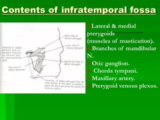
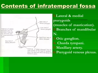
CONTENTS • Muscles- lateral & medial pterygoids, temporalis • Nerves- mandibular nerve & branches, chorda tympani nerve, Otic ganglion and connections • Vessels- maxillary artery & pterygoid venous plexus
LATERAL PTERYGOID MUSCLE • key muscle • Origin- 2 heads Upper- sphenoid Lower- lateral pterygoid plate • Insertion-pterygoid fovea, capsule & articular disc of TMJ.
LATERAL PTERYGOID • Nerve supply- anterior division of mandibular nerve • Actions- • Depression of mandible. • Protrusion of mandible - • Side- side chewing movements
RELATIONS • Superficial – • maxillary artery • Tendon of temporalis • Ramus of mandible • Masseter • deep – • mandibular nerve and branches
Relations • Upper border –deep temporal and masseteric nerves • Lower border -lingual & inferior alveolar nerve • Sphenomandibular ligament • Middle meningeal artery.
MEDIAL PTERYGOID • Origin-2 heads Deep- lateral pterygoid plate Superficial- tuberosity of maxilla & palatine bone • Insertion- medial surface of ramus & angle of mandible
MEDIAL PTERYGOID • Nerve supply- branch from trunk of mandibular nerve • Actions- • Elevation of mandible • Protrusion of mandible • Side- side chewing movements
RELATIONS • Lateral surface – • Ramus of mandible • Lateral pterygoid muscle • Sphenomandibular ligament. • Maxillary artery • Inferior alveolar vessels & nerve • Lingual nerve • Medial surface- • Tensor veli palatini • Superior constrictor of pharynx • Styloglossus and stylopharyngeus
Masseter • Origin:Zygomatic Arch. • Insertion:Lateral surface of the ramus of the mandible. • Action:Elevation of the mandible combined with protraction
Temporalis Origin: temporal fossa, temporal fascia . Insertion: coronoid process of the mandible. Action: • Elevates the mandible • Retraction – posterior fibers
MANDIBULAR NERVE • Largest of 3 divisions of trigeminal nerve • Mixed nerve - sensory + motor root present • Sensory - trigeminal ganglion • Motor - pons • Emerge through Foramen ovale
MANDIBULAR NERVE Trigeminal ganglion pons Sensory root Motor root Middle cranial fossa Foramen ovale unite Infratemporalfossa Trunk of the mandibular nerve Otic ganglion & middle meningeal artery lies behind the trunk. Medially-tensor velipalatini. Laterally-lateral pterygoid.
BRANCHES: MANDIBULAR ERVE TRUNK MENINGEAL BRANCH NERVE TO THE MEDIAL PTERYGOID ANTERIOR POSTERIOR PREDOMINANTLY MOTOR PREDOMINANTLY SENSORY MASSETERIC NERVE AURICULO-TEMPORAL NERVE DEEP TEMPORAL NERVES INFERIOR ALVEOLAR NERVE NERVE TO THE LATERAL PTERYGOID LINGUAL NERVE BUCCAL NERVE
MENINGEAL BRANCH(NERVUS SPINOSUS) FORAMEN SPINOSUM MIDDLE MENINGEAL ARTERY
MASSETERIC NERVE MASSETERIC NERVE
MASSETERIC NERVE TEMPORALIS LATERAL PTERYGOID MASSETERIC NERVE AND ARTERY INSERTION OF MASSETER MUSCLE
DEEP TEMPORAL NERVES POSTERIOR DEEP TEMPORAL NERVE ANTERIOR DEEP TEMPORAL NERVE TEMPORALIS
NERVE TO LATERAL PTERYGOID LATERAL PTERYGOID MUSCLE
BUCCAL NERVE BUT NOT BUCCINATOR MUSCLE BUCCAL NERVE Skin of the mucous membrane of the cheek and gum of lower jaw opposite molar and premolar BUCCINATOR BUCCAL NERVE BUCCAL NERVE
AURICULOTEMPORAL NERVE 1.AURICULAR BRANCHES 1.SKIN OF THE TRAGUS 2.UPPER PART OF THE AURICLE 3.ROOF AND ANTERIOR WALL OF THE EXTERNAL ACOUSTIC MEATUS 4.CUTICULAR LAYER OF TYMPANIC MEMBRANE. 2.SUPERFICIAL TEMPORAL BRANCHES SKIN OF THE TEMPLE 3.ARTICULAR BRANCHES TEMPOROMANDIBULAR JOINT POSTERIOR ROOT OF THE ZYGOMA SUPERFICIAL TEMPORAL ARTERY AURICULOTEMPORAL NERVE SPHENO-MANDIBULAR LIGAMENT LATERAL PTERYGOID MUSCLE MIDDLE MENINGEAL ARTERY
INFERIOR ALVEOLAR NERVE MYLOHYOID NERVE LATERAL PTERYGOID MUSCLE MYLOHYOID SUBMANDIBULAR GLAND SPHENOMANDIBULAR LIGAMENT INFERIOR ALVEOLAR ARTERY
LINGUAL NERVE TENSOR VELI PALATINI MUSCLE . . . . . . . INFERIOR ALVEOLAR NERVE . LATERAL PTERYGOID MUSCLE CHORDA TYMPANI NERVE MEDIAL PTERYGOID MUSCLE
LINGUAL NERVE THIRD MOLAR TOOTH MYLOHYOID MUSCLE STYLOGLOSSUSMUSCLE HYOGLOSSUS SUBMANDIBULAR GANGLION HYPOGLOSSAL NERVE SUBMANDIBULAR DUCT
LINGUAL NERVE • DISTRIBUTION: • GENERAL SENSE TO PRE-SULCAL PART OF THE • TONGUE • TO THE FLOOR OF THE MOUTH • MANDIBULAR GUM.
LINGUAL NERVE COMMUNICATIONS • COMMUNICATES WITH: • CHORDA TYMPANI NERVE • SUBMANDIBULAR GANGLION. • SECRETO-MOTOR FIBRES TO : • SUBMANDIBULAR GLAND • SUBLINGUAL GLAND. • TASTE SENSATION FROM: • ANTERIOR TWO THIRD OF THE TONGUE.
CHORDA TYMPANI NERVE • Branch of facial nerve • Arises – • 6mm above stylomastoid foramen. • Medial to spine of sphenoid • Joins lingual nerve at acute angle
OTIC GANGLION • Small,oval,parasympathetic ganglion. • 2-3 mm in size. • Situation – infratemporal fossa. • Topographically – mandibular nerve. • Functionally– glossopharyngeal nerve. • SITUATION: • Immediately below the foramen ovale. • Medial to the trunk of the mandibular • nerve. • Lateral to the tensor veli palatini muscle. • Infront of middle meningeal artery. • Behind the medial pterygoid muscle.
CONNECTIONS AND BRANCHES OF OTIC GANGLION • PARASYMPATHETIC ROOT Preganglionic fibers Inferior salivatory nucleus Tympanic branch of the CN IX Tympanic plexus Lesser petrosal nerve ganglion relay
CONNECTIONS AND BRANCHES OF OTIC GANGLION Post ganglionic fibers Ganglion Auriculo temporal nerve Secretomotorfibres to the parotid gland.
CONNECTIONS AND BRANCHES OF OTIC GANGLION • Sympathetic root Superior cervical ganglion Plexus around the middle meningeal artery Ganglion Auriculotemporal nerve Vasomotor supply to the parotid gland
Sensory branches • Auriculo temporal nerve • Somatic branches • Nerve to medial pterygoid • Supplies • Medial pterygoid • Tensor veli palatini • Tensor tympani