Comparative Evaluation of Genetic Markers in Primary RCC
This study compared microsatellite markers, AP-PCR, and CGH in primary RCC samples, highlighting specific genetic alterations in different histological types of renal cell carcinoma. The findings shed light on the genetic landscape of RCC.

Comparative Evaluation of Genetic Markers in Primary RCC
E N D
Presentation Transcript
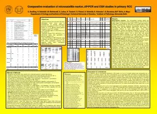
Comparative evaluation of microsatellite marker, AP-PCR and CGH studies in primary RCC • C. Hoefling, H. Schmidt*, M. Meinhardt#, A. Lohse, H. Taubert*, S. Füssel, U. Schmidt, K. Schuster*, G. Baretton#, M.P. Wirth, A. Meye • Department of Urology and Institute of Pathology#, Technical University of Dresden, *Institute of Pathology, University Halle Results Microsatellite Analysis. The microsatellite markers Bat25 and Bat26 were excluded from further studies because, within the first 40 tumor cases, no alterations were found. PCR´s were performed for all 99 RCC samples with the remaining microsatellite markers. Seven primary RCC samples displayed microsatellite alterations (7%) including LOH and shift patterns (MSI) (Fig1). The number of aberrations differed for each case (Tab3). Three cases (#34,#74,#100) showed microsatellite alterations at one locus, two samples (#13, #63) at 2 loci and also 2 samples (#40,#41) at 4/9 studied microsatellite markers. Five of seven RCC with altered microsatellite patterns belonged to the clear cell type, one was a papillary and one a chromophobe RCC, respectively (Tab3). Within the other 92 RCC, no abnormalities were identified (Tab2). Alu I/ AP-PCR Analysis. In only one (#13) of the 99 tumor samples genomic instability could be detected by AP-PCR approach (Fig 2). This result was confirmed in two independent experiments. CGH Analysis. In order to obtain a genomewide survey of tumor-associated alterations, CGH was performed additionally on RCC samples that were positive (n=6) or negative (n=4) for microsatellite alterations. Changes in copy number of DNA sequences were detected in five cases (#13, #34, #63, #74, #95) whereby four of the five samples had been already classified as positive for microsatellite alterations. #95 samples (no microsatellite alterations) also displayed genomic changes. Tumor samples with genomic imbalances showed a mean number of four alterations per sample (range 2-7). The most frequently affected chromosomal regions are summarized in Figure 3. Table 3 shows a detailed description of copy number changes of the examined RCC. As shown in Figure 3, nine chromosomal gains within six chromosomes were observed. The minimal common regions of gains are 5q14-q23 (cases #13, #95) and 7q11.1-q32 (cases #74, #95). Losses were more frequent than gains. Altogether, 11 DNA losses were observed on six chromosomes. Minimal common regions of DNA loss were 3p14-pter for three cases (#13, #34, #63), 13q13-qter (#13, #63), and 14q22-qter (#34, #63). • Objectives • - genetic analyses demonstrated that each histological renal tumor type is characterized by specific alterations extensive investigation of RCC by CGH, Junker et al. (3)defined typical genetic changes on each RCC subtype • CGH provides data for construction of a tree model (4) • arbitrarily primed PCR (AP-PCR) is an alternative assay that generates a genomic fingerprint (5) • here, the findings of MSI analyses using 9 markers with those detected in the AP-PCR were compared; positive and negative cases were investigated additionally by CGH Table 2 Primers Table 1: Clinico-pathological data of the 99 patients with renal cancers Table 3 Figure 2 Figure 1 Legends of Figures Fig1: Microsatellite alterations detected in renal cancers. Altered patterns indicate MSI and LOH, respectively. ; Fig2: AP-PCR analysis - Alterations in sample #13 are marked with a box.; Fig3: Summary of gains and losses of DNA sequence copy number in RCC samples analyzed by CGH. Losses are shown on the left, gains on the right side. Figure 3 Discussion & Conclusions The detection rate of microsatellite alterations (7/99 cases) for 6 out of 9 tested markers was comparably low. In contrast to colorectal cancers (16), RCC tumorigenesis is not associated either with Bat25 nor Bat26 alterations. Furthermore, no genetic abnormalities were revealed for REN (1q32). These results could be in line with two recent studies describing that the occurrence of mutations in mismatch repair genes and the down-regulation of mismatch repair gene expression is uncommon in RCC-derived cell lines and tissues (17,18). The p53 gene and the myc gene were studied by the microsatellite markers TP53Alu- and Myc-L1-PCR’s revealing three positive out of 99 tested samples in each locus. Previous reports showed genetic changes in RCC at 17p ranging up to 35% (19,20). This suggests, that genetic alterations of the p53 gene harbored at 17p reflect accumulating events rather than a locus for an initial events (19). The VHL region displayed LOH in 21% of the investigated tumor samples (D3S4260, D3S1560), respectively (23). However, in the RCC study by Velickovic et al. 58% of the evaluated tumor samples were diagnosed as tumors of higher stages (T3,T4). In comparison, in our patients cohort, only 19% of the investigated RCC were stage T3 tumors, whereas no stage T4 RCC were investigated. Other authors (24) also described the lack of MSI for RCC in younger Americans. We identified in only one (#13) out of 99 cases a shift in AP-PCR band pattern (Fig. 2). This tumor sample showed also in the MSI and CGH investigations (Table 1). Because of the overall low frequency of detected abnormalities the AP-PCR approach is not suitable as a fingerprint for the identification of genetic instability in RCC. Within these few cases the most frequent genetic change was DNA sequence copy number loss within chromosome 3p as described from others (26-28). Two samples of clear cell-type RCC (#13,#34) displayed losses within the chromosomes 3, 4, 8, 13 and 14 as well as a gain of DNA sequence on chromosome 5. These findings also correlate with previous studies (28-30). Focussing on alterations detected by microsatellite analysis, AP-PCR and CGH investigation, no significant correlation could be found between patients with alterations and an expected prognosis (Table 3). In summary, comparing three different methods to detect genetic alterations in primary RCC, we found in five out of seven tumor samples with microsatellite alterations an aberration at 3p. In two cases aberrations at 3p were detected by CGH as well. AP-PCR appears less informative at investigating RCC. • Material & Methods • 99 patients with primary renal cancers who underwent partial or radical nephrectomy • samples of tumor and normal tissue were snap-frozen in liquid nitrogen and stored at -80 °C • tumor staging according to the Mainz classification (6) and the 2003 TNM (UICC): • 63 clear cell, 21 chromophobe, 12 papillary, 1 duct carcinoma, 1 oncocytoma, 1 sarcomatoid • mean age at diagnosis was 63 years (range 32-89) (Table 1) • - genomic DNA isolation from 50µm tissue sections (proteinase K digestion, salt extraction (7) • DNA isolation for control blood samples and for CGH: Ficoll gradient centrifugation, salt extraction • - Microsatellite investigation: 9 microsatellite loci (Table 2) • amplifications: 0.5µM primer, 100ng DNA, 0.1-0.2mM dNTPs, 1-2.5 mM MgCl2, 2.5U Taq, 10ng BSA • PCR conditions according to the original protocols (Table 2): 94°C for 5min, samples, 35 or 40 cycles of 94°C/1min, 53-70°C/1min, 72°C/1 min, & 72°C/8min • products were resuspended in a formamide buffer, 94°C/5min, loaded on a 6.7% PAA gel • silver staining method (Sammoris et al. (8)); alterations in markers were defined as MSI or LOH • AP-PCR generates genomic DNA-fingerprint using the presence of Alu sequences (5) • Alu I DNA restriction (500ng DNA, 6U Alu I), at 37°C for 16h, AP-specific primer from (4) • 200ng of Alu I digested DNA, PCR with 0.5µM AP-primer, 0.2 mM dNTPs, 5mM MgCl2, 5U Taq • CGH: 6 samples positive for microsatellite alterations & 4 negative samples (Table 3) • reference DNA was prepared from blood of healthy probands • CGH analysis according to standard protocols (9) with some modifications (10): • hybridization mixture: 1-3µg DNA, 40µg Cot-1 human DNA, 50µg salmon sperm DNA; DNA was denatured at 78°C for 7min, pre-annealed at 42°C for 30min; lymphocyte metaphase were denatured at 72°C for 2min in 70% formamide; hybridization at 42°C for 3 days • fluorescence microscopy & ISIS digital image system (10), images from 10 metaphase cells: regions with signal ratio >1.15 were considered to be gains, regions with ratio <0.85 were considerd as losses References 1. Pavlovich CP, et al., Urol Clin North Am 30: 437-54, 2003. 2. Moch H, Mihatsch MJ, Virchows Arch 441: 320-327, 2002. 3. Junker K, et al., Recent Results Cancer Res 162: 169-75, 2003. 4. Jiang F, et al. Cancer Res 60: 6503-9, 2000. 5. Sood AK, Buller RE, Oncogene 13: 2499-2504, 1996. 6. Thoenes W, Störkel S, Urologe A 30: W41-50, 1991. 7. Lahiri DK, Nurnberger JI, Nucleid Acids Res 19: 5444, 1991. 8. Sammoris DW, et al. Electrophoresis 2: 135-41, 1981. 9. Kallioniemi OP, et al. Genes Chromosomes Cancer 10: 231-243, 1994. 10. Schmidt H, et al., Genes Chromosomes Cancer 25: 205-211, 1999. 11. Jass JR, et al., Lancet 346: 1200-1, 1995. 12. Papadopoulos N, et al, Science 268: 1915-1917, 1995. 13. Mazela TP, et al., Hum Mol Genet 1: 217, 1992. 14. Futureal P, et al., Nucleic Acids Res 19: 6977, 1991. 15. Edwards A, et al., Genomics 12: 241-253, 1992. 16. Nash GM, et al., J Clin Oncol 21: 3105-12, 2003. 17. Leach FS, et al., Cancer Biol Ther 1: 530-6, 2002. 18. Deguchi M, et al., J Urol 169: 2365-71, 2003. 19. Brauch H, et al., World J Urol 12: 162-168, 1994. 20. Uchida T, et al., J Urol 150: 1298-1301, 1993. 21. Dietmaier W, et al., Cancer Res 57:4749-56, 1997. 22. Bugert P, et al., Genes Chromosomes Cancer 20: 9-15, 1997. 23. Velickovic M, et al., Cancer Res 59: 1323-1326, 1999. 24. Kanomata N, et al., Cancer Genet Cytogenet 101: 123-7, 1998. 25. Uchida T, et al., Cancer Res 54:3682-5, 1994. 26. Zbar B, et al., Nature 327: 721-727, 1987. 27. Morita R, et al., Cancer Res 51: 820-823, 1991. 28. Presti J, et al., Cancer Res 53: 5780-5783, 1993. 29. Moch H, et al., Cancer Res 56: 27-30, 1996. 30. Reutzel D, et al., Cytogenet Cell Genet 93: 221-227, 2001. 31. Bentz M, et al., Cytogenet Cell Genet 75: 17-21, 1996. 32. Jiang F, et al., Am J Pathol 153: 1467-1473, 1998. 33. Speicher M, et al., Am J Pathol 145: 356-364, 1994. 34. Jiang F, et al., J Pathol 185: 382-8, 1998. 35. Moch M, et al., Cancer 89: 604-614, 2000. 36. Nimer S, Golde D, Blood 70: 1705-1712, 1987. 37. Kovacs G, Frisch S, Cancer Res 49: 651-659, 1989. 38. Kovacs G, Histopathology 22: 1-8, 1993. 39. Gonzalgo ML, et al., Clin Cancer Res 8: 1878-81, 2002. 40. Gyapay G, et al., Nat Genet 7: 246-339, 1994. 41. Li H, et al., Hum Mol Genet 2: 1326, 1993. 42. Weissenbach J, et al. Nature 359, 1992.