Download
1 / 77
800 likes | 841 Views
Explore how our bodies combat infections through white blood cell function. Learn how leukocytes work to prevent diseases, the differentiation of stem cells, and the process of phagocytosis.

E N D
Our body has a special system for fightingagainst different infectious and toxic agents; • blood leukocytes and tissue cells derived from leukocytes; they work together in two ways to prevent disease: (1) by actually destroying invading bacteria or viruses by phagocytosis, and (2) by forming antibodies and sensitized lymphocytes, one or both of which may destroy or inactivate the invader.
The leukocytes (white blood cells)- mobile units of the body’s protective system. • are formed partially in the bone marrow (granulocytes and monocytes and a few lymphocytes) and partially in the lymph tissue (lymphocytes and plasma cells). • are transported in the blood to different parts of the body where they are needed.
Six types of WBCs are normally present in the blood: polymorphonuclear neutrophils, polymorphonuclear eosinophils, polymorphonuclear basophils, monocytes, lymphocytes. • The polymorphonuclear cells, all have a granular appearance, for which reason they are called granulocytes, or, in clinical terminology,“polys,” because of the multiple nuclei.
Normal values (white blood cells): 4500÷10 000/mm ³ • Neutrophils 60-65% • Eosinophils 1-3% • Basophils 0.15-0.5% • Monocytes 4-8% • Lymphocytes 25-35% • Band neutrophils = maximum 10% from segmented neutrophils = 2-6%
The Pluripotential hematopoietic stem cell are differentiatedinto the different types of committed stem cells • Aside from those cells committed to form red blood cells, two major lineages of white blood cells are formed, the myelocytic and the lymphocytic lineages. • the myelocytic lineage, beginning with the myeloblast; the lymphocytic lineage, beginning with the lymphoblast.
The life of the granulocytes after being released from the bone marrow is normally 4 to 8 hours circulating in the blood and another 4 to 5 days in tissues where they are needed. • In case of tissue infection, this total life span is often shortened to only a few hours • The monocytes also have a short transit time, 10 -20 hours in the blood, before wandering through the capillary membranes into the tissues.
The neutrophils are mature cells that can attack and destroy bacteria even in the circulating blood. • The tissue macrophages begin life as blood monocytes; once they enter the tissues, they begin to swell—sometimes increasing their diameters as much as fivefold—to as great as 60 to 80 micrometers. These cells are now called macrophages, and they are extremely capable of combating intratissue disease agents. • Neutrophils and monocytes can squeeze through the pores of the blood capillaries by diapedesis.
Both neutrophils and macrophages can move through the tissues by ameboid motion. Some cells move at velocities as great as 40 mm/min, a distance as great as their own length each minute. • Many different chemical substances in the tissues cause both neutrophils and macrophages to move toward the source of the chemical. This phenomenon, is known as chemotaxis. When a tissue becomes inflamed, at least a dozen different products are formed that can cause chemotaxis toward the inflamed area.
They include (1) some of the bacterial or viral toxins, (2) degenerative products of the inflamed tissues themselves, (3) several reaction products of the “complement complex” activated in inflamed tissues, and (4) several reaction products caused by plasma clotting in the inflamed area, as well as other substances. Chemotaxis depends on the concentration gradient of the chemotactic substance. The concentration is greatest near the source, which directs the unidirectional movement of the white cells.
The most important function of the neutrophils and macrophages is phagocytosis=cellular ingestion of the offending agent. • Phagocytes must be selective of the material that is phagocytized; otherwise, normal cells and structures of the body might be ingested. Whether phagocytosis will occur depends especially on three selective procedures: • smooth surfaces, which resist phagocytosis. But if the surface is rough, the likelihood of phagocytosis is increased. • most natural substances of the body have protective protein coats that repel the phagocytes. Conversely, most dead tissues and foreign particles have no protective coats, which makes them subject to phagocytosis.
Third, the immune system of the body develops antibodies against infectious agents such as bacteria. The antibodies then adhere to the bacterial membranes and thereby make the bacteria especially susceptible to phagocytosis. • To do this, the antibody molecule also combines with the C3 product of the complement cascade (an additional part of the immunesystem). • The C3 molecules, in turn, attach to receptors on the phagocyte membrane, thus initiating phagocytosis. This selection and phagocytosis process is called opsonization.
The neutrophils entering the tissues are already mature cells that can immediately begin phagocytosis: • first attaches itself to the particle and then projects pseudopodia in all directions around the particle. • the pseudopodia meet one another on the opposite side and fuse.This creates an enclosed chamber that contains the phagocytized particle. • then the chamber invaginates to the inside of the cytoplasmic cavity and breaks away from the outer cell membrane to form a free-floating phagocytic vesicle (also called a phagosome) inside the cytoplasm.
Once a foreign particle has been phagocytized, lysosomes and other cytoplasmic granules in the neutrophil or macrophage immediately come in contact with the phagocytic vesicle, and their membranes fuse, thereby dumping many digestive enzymes and bactericidal agents into the vesicle. • Both neutrophils and macrophages contain an abundance of lysosomes filled with proteolytic enzymes for digesting bacteria and other foreign protein matter, large amounts of lipases, which digest the thick lipid membranes possessed by some bacteria such as the tuberculosis bacillus.
In addition, neutrophils and macrophages contain bactericidal agents that kill most bacteria even when the lysosomal enzymes fail to digest them. • Much of the killing effect results from several powerful oxidizing agents formed by enzymes in the membrane of the phagosome or by a special organelle called the peroxisome. • These oxidizing agents include large quantities of superoxide (O2–) hydrogen peroxide (H2O2), and hydroxyl ions (–OH–), all of which are lethal to most bacteria, even in small quantities
One of the lysosomal enzymes, myeloperoxidase, catalyzes the reaction between H2O2 and chloride ions to form hypochlorite, which is exceedingly bactericidal. • Some bacteria, however, notably the tuberculosis bacillus, have coats that are resistant to lysosomal digestion and also secrete substances that partially resist the killing effects of the neutrophils and macrophages. These bacteria are responsible for many of the chronic diseases, an example of which is tuberculosis.
Afterenteringthetissuesandbecomingmacrophages, anotherlargeportion of monocytesbecomesattachedtothetissuesandremainsattached for months or evenyearsuntilthey are called on toperformspecific local protective functions. • Thus,the body has a widespread “monocyte-macrophagesystem” in virtuallyalltissueareas. • The total combination of monocytes, mobile macrophages, fixedtissuemacrophages, and a fewspecializedendothelialcells in the bone marrow, spleen, andlymphnodesiscalledthereticuloendothelialsystem.
Inflammation • When tissue injury occurs, whether caused by bacteria, trauma, chemicals, heat, or any other phenomenon, multiple substances are released by the injured tissues and cause dramatic secondary changes in the surrounding uninjured tissues. • This entire complex of tissue changes is called inflammation.
Inflammation is characterized by (1) vasodilation of the local bloodvessels(excess local blood flow); (2) increased permeability of the capillaries, (leakage of large quantities of fluid into the interstitial spaces); (3) often clotting of the fluid in the interstitial spaces because of excessive amounts of fibrinogen and other proteins leaking from the capillaries; (4) migration of large numbers of granulocytes and monocytes into the tissue; and (5) swelling of the tissue cells.
Some of themanytissueproductsthatcausethesereactions are histamine, bradykinin, serotonin, prostaglandins, severaldifferentreactionproducts of the complement system, reactionproducts of thebloodclottingsystem, and multiple substancescalledlymphokinesthat are releasedbysensitizedT cells. • Several of thesesubstancesstrongly activate themacrophagesystem, andwithin a fewhours, themacrophagesbegintodevourthedestroyedtissues. But at times, themacrophagesalsofurtherinjurethestill-living tissuecells.
Within minutes after inflammation begins, the macrophages already present in the tissues immediately begin their phagocytic actions. • The first effect is rapid enlargement of each of these cells. • Next, many of the previously macrophages break loose from their attachments and become mobile, forming the first line of defense against infection during the first hour or so. • Within the first hour or so after inflammation begins, large numbers of neutrophils begin to invade the inflamed area from the blood. • This is caused by products from the inflamed tissues that initiate the following reactions:
(1) alteration the inside surface of the capillary endothelium, causing neutrophils to stick to the capillary walls in the inflamed area. This effect is called margination; (2) cause the intercellular attachments between the endothelial cells of the capillaries and small venules to loosen, allowing openings large enough for neutrophils to pass by diapedesis directly from the blood into the tissue spaces. (3) Other products of inflammation then cause chemotaxis of the neutrophils toward the injured tissues.
Thus, within several hours after tissue damage begins, the area becomes well supplied with neutrophils. Because the blood neutrophils are already mature cells,they are ready to immediately begin their scavenger functions for killing bacteria and removing foreign matter.
Also within a few hours after the onset of acute, severe inflammation, the number of neutrophils in the blood sometimes increases fourfold to five- fold—from a normal of 4000 to 5000 to 15,000 to 25,000 neutrophils per microliter. This is called neutrophilia, which means an increase in the number of neutrophils in the blood. • Neutrophilia is caused by products of inflammation that enter the blood stream, are transported to the bone marrow, and there act on the stored neutrophils of the marrow to mobilize these into the circulating blood.
The number of monocytes in the circulating blood is low: also, the storage pool of monocytes in the bone marrow is much less than that of neutrophils. • Therefore, the buildup of macrophages in the inflamed tissue area is much slower than that of neutrophils, requiring several days to become effective. • Even after invading the inflamed tissue, monocytes are still immature cells, requiring 8 hours or more to swell to much larger sizes and develop tremendous quantities of lysosomes; only then do they acquire the full capacity of tissue macrophages for phagocytosis.
The next line of defense is greatly increased production of both granulocytes and monocytes by the bone marrow. This results from stimulation of the granulocytic and monocytic progenitor cells of the marrow. • It takes 3 to 4 days before newly formed granulocytes and monocytes reach the stage of leaving the bone marrow. If the stimulus from the inflamed tissue continues,the bone marrow can continue to produce these cells in tremendous quantities for months and even years, sometimes at a rate 20 to 50 times normal.
When neutrophils and macrophages engulf large numbers of bacteria and necrotic tissue, essentially all the neutrophils and many, if not most, of the macrophages eventually die. • After several days, a cavity is often excavated in the inflamed tissues that contains varying portions of necrotic tissue, dead neutrophils, dead macrophages, and tissue fluid. • This mixture is known as pus. After the infection has been suppressed, the dead cells and necrotic tissue in the pus gradually autolyze over a period of days, and the end products are eventually absorbed into the surrounding tissues and lymph until most of the evidence of tissue damage is gone.
Eosinophils • The eosinophils normally range 1-3% • are weak phagocytes, and they exhibit chemotaxis • are often produced in large numbers in people with parasitic infections, and they migrate in large numbers into tissues diseased by parasites. • Although most parasites are too large to be phagocytized by eosinophils or any other phagocytic cells, eosinophils attach themselves to the parasites by way of special surface molecules and release sub stances that kill many of the parasites.
Eosinophils attach themselves to the juvenile forms of the parasite and kill many of them. • They do so in several ways: (1) by releasing hydrolytic enzymes from their granules, which are modified lysosomes; (2) probably by also releasing highly reactive forms of oxygen that are especially lethal to parasites;and (3) by releasing from the granules a highly larvacidal polypeptide called major basic protein
Eosinophils also have a special propensity to collect in tissues in which allergic reactions occur, such as in the peribronchial tissues of the lungs in people with asthma and in the skin after allergic skin reactions. • This is caused at least partly by the fact that many mast cells and basophils participate in allergic reactions (they release an eosinophil chemotactic factor that causes eosinophils to migrate toward the inflamed allergic tissue.
Basophils • The basophils in the circulating blood are similar to the large tissue mast cells located immediately outside many of the capillaries in the body. Both mast cells and basophils liberate heparin into the blood, a substance that can prevent blood coagulation. • The mast cells and basophils also release histamine, as well as smaller quantities of bradykinin and serotonin during inflammation. • The mast cells and basophils play an exceedingly important role in some types of allergic reactions
A clinical condition known as leukopenia occasionally occurs in which the bone marrow produces very few white blood cells, leaving the body unprotected against many bacteria and other agents that might invade the tissues. • Normally, the human body lives in symbiosis with many bacteria, because all the mucous membranes of the body are constantly exposed to large numbers of bacteria
Irradiation of the body by x-rays or gamma rays, or exposure to drugs and chemicals that contain benzene or anthracene nuclei, is likely to cause aplasia of the bone marrow. • Some common drugs, such as chloramphenicol, thiouracil (used to treat thyrotoxicosis), and even various barbiturate hypnotics, on very rare occasions cause leukopenia, thus setting off the entire infectious sequence of this malady. • After moderate irradiation injury to the bone marrow, some stem cells, myeloblasts, and hemocyto blasts may remain undestroyed in the marrow and are capable of regenerating the bone marrow, provided sufficient time is available.
Uncontrolled production of white blood cells can be caused by cancerous mutation of a myelogenous or lymphogenous cell. • This causes leukemia, which is usually characterized by greatly increased numbers of abnormal white blood cells in the circulating blood. • Are divided into two general types: lymphocytic leukemias and myelogenous leukemias.
The first effect of leukemia is metastatic growth of leukemic cells in abnormal areas of the body. • Leukemic cells from the bone marrow may reproduce so greatly that they invade the surrounding bone, causing pain and, eventually, a tendency for bones to fracture easily. • Almost all leukemias eventually spread to the spleen, lymph nodes, liver, and other vascular regions, regardless of whether the origin of the leukemia is in the bone marrow or the lymph nodes. • Common effects in leukemia are the development of infection, severe anemia, and a bleeding tendency caused by thrombocytopenia (lack of platelets).
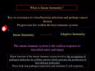
Innate Immunity • The human body has the ability to resist almost all types of organisms or toxins that tend to damage the tissues and organs = immunity. Much of immunity is acquired immunity that does not develop until after the body is first attacked by a bacterium, virus, or toxin, often requiring weeks or months to develop it. • An additional portion of immunity results from general processes, rather than from processes directed at specific disease organisms. This is called innate immunity.
Innate immunity includes the following: 1. Phagocytosis of bacteria and other invaders by white blood cells and cells of the tissue macrophage system. 2. Destruction of swallowed organisms by the acid secretions of the stomach and the digestive enzymes. 3. Resistance of the skin to invasion by organisms.
4. Presence in the blood of certain chemical compounds that attach to foreign organisms or toxins and destroy them (1) lysozyme, a mucolytic polysaccharide that attacks bacteria and causes them to dissolute; (2) basic polypeptides, which react with and inactivate certain types of gram-positive bacteria; (3) the complement complex (system of about 20 proteins that can be activated in various ways to destroy bacteria) and (4) natural killer lymphocytes that can recognize and destroy foreign cells, tumor cells, and even some infected cells
Acquired (Adaptive) Immunity • the human body has the ability to develop extremely powerful specific immunity against individual invading agents such as lethalbacteria,viruses,toxins,and even foreign tissues from other animals = acquired or adaptive immunity. • Acquired immunity is caused by a special immune system that forms antibodies and/or activated lymphocytes that attack and destroy the specific invading organism or toxin.
The body develops circulating antibodies, which are globulin molecules in the blood plasma that are capable of attacking the invading agent. This type of immunity is called humoral immunity or B-cell immunity (because B lymphocytes produce the antibodies). • The second type of acquired immunity is achieved through the formation of large numbers of activated T lymphocytes that are specifically crafted in the lymph nodes to destroy the foreign agent. This type of immunity is called cell-mediated immunity or T-cell immunity (because the activated lymphocytes are T lymphocytes).
Because acquired immunity does not develop until after invasion by a foreign organism or toxin, it is clear that the body must have some mechanism for recognizing this invasion. • Each toxin or each type of organism almost always contains one or more specific chemical compounds in its makeup that are different from all other compounds. In general, these are proteins or large polysaccharides, and it is they that initiate the acquired immunity and are called antigens(antibody generations). • The process of antigenicity usually depends on regularly recurring molecular groups, called epitopes, on the surface of the large molecule. This also explains why proteins and large polysaccharides are almost always antigenic, because both of these have this stereochemical characteristic.
Lymphocytes are distinctly divided into two major populations. One of the populations, • the T lymphocytes, is responsible for forming the activated lymphocytes that provide “cell-mediated” immunity, and • the B lymphocytes, is responsible for forming antibodies that provide “humoral” immunity. • Both types of lymphocytes are derived originally in the embryo from pluripotent hematopoietic stem cells that form lymphocytes as one of their most important offspring as they differentiate.
Almost all of the lymphocytes that are formed eventually end up in the lymphoid tissue, but before doing so, they are further differentiated or “preprocessed”in the following ways. • The lymphocytes that are destined to eventually form activated T lymphocytes first migrate to and are preprocessed in the thymus gland, and thus they are called “T” lymphocytes to designate the role of the thymus. • The B lymphocytes that are destined to form antibodies—are preprocessed in the liver during midfetal life and in the bone marrow in late fetal life and after birth.
The T lymphocytes, afterorigination in the bone marrow, first migrate tothethymus gland. Here they divide rapidlyand at the same timedevelop extreme diversity for reactingagainstdifferentspecificantigens.Thatis, onethymiclymphocytedevelopsspecificreactivityagainstone antigen. • Thiscontinuesuntilthere are thousands of differenttypes of thymiclymphocyteswithspecificreactivitiesagainstmanythousands of differentantigens. Thesedifferenttypes of preprocessed T lymphocytesnowleavethethymusandspreadbyway of thebloodthroughoutthe body tolodge in lymphoidtissueeverywhere
The thymusalsomakescertainthatany T lymphocytesleavingthethymuswillnotreactagainstproteins or otherantigensthat are present in thebody’sowntissues; otherwise, the T lymphocyteswouldbelethaltotheperson’sown body in only a fewdays. • The thymusselectswhich T lymphocyteswillbereleasedbyfirstmixingthemwithvirtuallyallthespecific “self-antigens” fromthebody’sowntissues. If a T lymphocytereacts, it isdestroyedandphagocytizedinstead of beingreleased.
B lymphocytes are different from T lymphocytes in two ways: • actively secrete antibodies that are the reactive agents.These agents are large protein molecules that are capable of combining with and destroying the antigenic substance. • have even greater diversity than the T lymphocytes, thus forming many millions of types of B-lymphocyte antibodies with different specific reactivities. After preprocessing, the B lymphocytes, like the T lymphocytes, migrate to lymphoid tissue throughout the body, where they lodge near but slightly removed from the T-lymphocyte areas.
Whenspecificantigens come in contact with T and B lymphocytes in thelymphoidtissue, certain of the T lymphocytesbecomeactivatedtoformactivated T cells, andcertain of the B lymphocytesbecomeactivatedtoformantibodies. • The activated T cellsandantibodies in turn reacthighlyspecificallyagainstthe particular types of antigensthatinitiatedtheirdevelopment. • The mechanism of thisspecificityisthefollowing.