Download
1 / 28
290 likes | 425 Views
Fluorescent Assay of a RING-type Ubiquitin Ligase. Mississippi State University November 2007. Our Team. Our Team - Students. Agricultural and Biological Engineering Graduate Robert Morris Undergraduate Lauren Beatty, Scott Tran, Joe Chen, Caleb Dulaney, Sam Pote, Karen Parks

E N D
Fluorescent Assay of a RING-type Ubiquitin Ligase Mississippi State University November 2007
Our Team - Students • Agricultural and Biological Engineering • Graduate • Robert Morris • Undergraduate • Lauren Beatty, Scott Tran, Joe Chen, Caleb Dulaney, Sam Pote, Karen Parks • Biochemistry • Graduate • Victor Ho • Undergraduate • James Kastrantas
Our Team - Professors • Agricultural and Biological Engineering • Dr. Filip To • Biochemistry • Dr. Din-Pow Ma • Electrical and Computer Engineering • Dr. Bob Reese • Chemical Engineering • Dr. Todd French
Present Areas of Interest • Departmental Goal • Discover lipid production pathways • Increase lipid synthesis in plants • Make biofuels more economically feasible • iGEM 2007 Goal • Design a construction that simplifies and expedites the confirmation of ubiquitin ligase activity
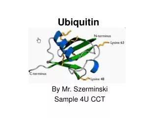
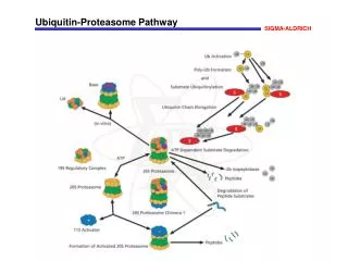
Ubiquitin Proteasome Pathway • The ubiquitination of target proteins for degradation requires the sequential activity of three enzymes: • E1, ubiquitin activating enzyme • E2, ubiquitin conjugating enzyme • E3, ubiquitin ligase
Methodology • A RING-type E3 gene, GhR1(1kb), was amplified by PCR and cloned into pGFPuv as an in-frame fusion at N-terminal with GFPuv. • Chemically competent (CaCl2) E. coli XL1-blue cells were transformed with recombinant plasmid pGFP/GhR1. • Transformed XL1-blue cells were grown to OD600 ~0.6 and lysed by sonnication.
Methodology • Cell lysate was added to ubiquitin and wheat germ extract, which provides reagents (E1, E2, ATP) for ubiquitination. • Reaction mixtures were analyzed by native PAGE. • Excitation of the fusions revealed the presence of E3 and ubiquitination on the gel.
Project Justification • Benefits: • Assays E3 ligase activity directly on native protein gel • Omits primary purification step, Western blot, and pull down assay • Saves time and resources
Desired Machine Function • Rapidly report ubiquitin ligase activity via green fluorescence
Problems Encountered Upon inspection of our construction plans involving the insertion of ubiquitin next to the Registry part J01095, the S/X restriction site created with the ligation of these two parts would code for the stop codon UAG.
Confirmation of Constructions • All constructions were sequenced using ABI 310 Prism Genetic Analyzer. • All sequences were confirmed to be correct and in frame with the GFP gene.
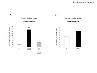
Results of First Construction LaneID 1 Protein Ladder 2 GhR1/GFP 3 GFP 4 GhR1/GFP/Ub 5 GFP/Ub 1 2 3 4 5 1 2 3 4 5 Ub – Ubiquitin
The GhR1-GFP fusion protein in lanes 2 and 4 had fluorescence. No multiple protein bands were detected in lane 4 after Western blotting with anti-ubiquitin, suggesting that ubiquitination did not occur. Analysis of First Construction
Results of Second Construction LaneID 1 Protein Ladder 2 Sh-GhR1/GFP 3 Fl- GhR1/GFP 4&5 GFP 1 2 3 4 5 Sh – short Fl – full
GFP without the fusion in lanes 4 and 5 showed fluorescence. There was no visible fluorescence in lanes 2 and 3, suggesting that the expression level of GhR1-GFP fusion was either too low or the fusion protein changed conformation with no fluorescence. Analysis of Second Construction
Conclusions • The first construction was successful with the observation of fluorescence from the GhR1-GFP fusion protein. • The first construction failed to detect poly-ubiquitin chains. • GFP adjacent to the RING domain (C-terminal) of GhR1 might block ubiquitination.
Conclusions • There was no expression of green fluorescence in the second construction. • Expression of the GhR1-GFP fusion was not observed in either the short or full length form of GhR1. • This could be the result of an alteration of protein structure from the fusion of GhR1 with GFP.
Future Work • Fusion of RFP with GhR1 • One single plasmid for both GFP-E3 and RFP-ubiquitin fusions • Discovery of other protein-protein interactions • Interactions controlling lipid synthesis
Future Work • Understanding of the regulation of lipid production will enable the design of new machines with predetermined lipid content • Higher lipid content in plant Higher energy value of biofuel • Lower undesired lipid levels Higher product value • May lead to understanding of other regulatory pathways (polysaccharides)
Acknowledgements • Advisors • Dr. Filip To, Dr. Din-Pow Ma • MSU Bagley College of Engineering • MSU College of Agriculture and Life Sciences • USDA Strategic Research Initiative • iGEM Staff