Normal Oral Mucosa
Normal Oral Mucosa. Oral mucosa differs considerably from the skin in: The oral mucosa varies in its firmness and texture. Generally, oral mucosa is more deeply colored (paler pink). The moist surface and the absence of appendages.

Normal Oral Mucosa
E N D
Presentation Transcript
Oral mucosa differs considerably from the skin in: • The oral mucosa varies in its firmness and texture. • Generally, oral mucosa is more deeply colored (paler pink). • The moist surface and the absence of appendages. • The surface of the oral mucosa tends to be smoother and have fewer folds and wrinkles than the skin.
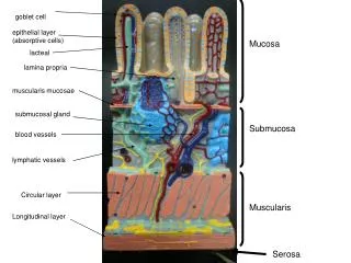
The oral cavity consists of two parts: an outer vestibule and the oral cavity proper. Classification basing on functional criteria, the oral mucosa may be divided into three major types: Masticatory mucosa (gingival and hard palate) Lining mucosa (oral side of the lip and cheek, floor of mouth, alveolar mucosa, ventrum of the tongue and soft palate) Specialized mucosa (dorsum of the tongue and taste buds, vermilion zone, Junctional epithelium, and lingual tonsils).Quantitatively, the larger part of the oral mucosa is represented by lining mucosa, amounting to 60% of the total area, with masticatory mucosa and specialized mucosa occupying smallest areas (25% and 15%, respectively).
Composition of MM Epithelium: 1.Basal cell layer 2.Prickle cell layer (stratum spinosum) 3.Granular cell layer (stratum granulosum) 4.Keratin layer
Oral epithelium • The oral epithelium is a stratified squamous epithelium consisting of cells tightly attached to each other and arranged in a number of distinct layers or strata. • The oral epithelium maintains its structural integrity by a process of continuous cell renewal in which cell produced by mitotic divisions in the deepest layers migrate to the surface to replace those that are shed. • The migrated cells undergo various structural modifications until they reach the surface. • These modifications are dependent on the process of keratinisation and vary according to the precise site of the mucosa involved. • The net result of this is the production of a surface layer of cells which are either fully, partially or non-keratinized. • The surface layer of cells are shed into the oral cavity at a rate dependent on the rate of mitosis at the basal layer.
The cells of the epithelium thus can be considered to consist of two functional populations: a progenitor population (the function of which is to divide and provide new cells) and maturing population (the cell of which continually undergo a process of differentiation or maturation to form a protective surface layer). • Variations in the extent of the keratinization process are shown in the epithelium of the oral mucosa. • In fully keratinized epithelium, the cubical cells migrate towards the surface and become more polyhedral. • They share intercellular attachments which have given the name “prickle cell layer” or “stratum spinosum” to this zone. • Cubical cells formed by mitosis at or near the basal layer. These intercellular junctions (desmosomes) are seen to be of much greater complexity by electron microscopy.
The desmosomes probably give strength to the epithelium by acting in a mechanical manner. • Hemidesmosomes, similar, one-sided structures, attach the plasma membrane of the basal cells to the lamina densa of the basement membrane complex. • The cells of the stratum spinosum begin to flatten and granular structures (keratohyalin granules) appear within them as they migrate to the surface. It is known that these granules, which give the characteristic appearance to the stratum granulosum in keratinized epithelia, are closely involved with the process of keratinization. • Finally, the epithelial cells loose their detailed inner structure at or near the surface. At this stage the desmosomes have degenerated and the flattened cells are eventually lost into the oral cavity. • This process applies only to fully keratinized epithelium and is usually referred to as orthokeratinization. • In other areas (buccal mucosa and the floor of the mouth) this process of keratinization does not occur. • However, in an intermediate form (parakeratotic epithelium) some of the chemical changes of keratinization occur in the superficial cells.
Melanocytes and oral pigmentation • The colour of the oral mucosa is the net result of a number of factors, one of which is pigmentation. • Two types of pigmentation occur: • endogenous, arising in the tissues from normal physiologic process, and exogenous, caused by foreign material introduced into the body locally (amalgam) or systemically (bismuth). • The endogenous pigments most commonly contributing to the colour of oral mucosa are melanin and haemoglobin. • Melanin is produced by specialized pigment cells, called melanocytes, situated in the basal layer of the oral epithelium and the epidermis. • Melanocytes arise embryologically from the neural crest ectoderm and enter the epithelium at about 11 week of gestation. • Melanin is synthesised in within the melanocytes as small structures called melanosomes, which are injected into the cytoplasm of adjacent keratinocytes by the dendritic processes of melanocytes. • Melanin granules can be identified under the light microscope.
Lightly and darkly pigmented individuals have the same number of melanocytes in any given region in oral mucosa or skin; • The degree of pigmentation is affected by: • The size and degree of branching of melanocytes (rather than by the number of cells). • The size of the melanosomes. • The number and degree of dispersion of melanosomes. • The degree of melanisation of the melanosomes. • Degradation rate of the pigment. • The regions of the oral mucosa where melanin pigmentation is seen most commonly clinically are the gingiva, buccal mucosa, hard palate and tongue. • Light-skinned persons rarely show any oral melanin pigmentation.
Langerhans' cells • Langerhans' cells can appear in the epithelium at the same time as, or just before, the melanocytes, and they may be capable of limited division within the epithelium. • Unlike melanocytes, they move in and out of the epithelium • Their source is the bone marrow. • Evidence suggests that langerhans' cells have an immunologic function, recognising and processing antigenic material that enters the epithelium from the external environment and presenting it to T lymphocytes. • They can migrate from epithelium to regional lymph nodes.
Langerhans Cells • Functions: • They play an important role in contact hypersensitivity reactions of the skin. • They have a role in anti-tumour immunity. • They also have a role in graft rejection. • They react as propagators of HIV-1 transmission to T cells.
Merkel Cells • Found in the basal cell layer. • Close to nerve fibres. • Acts as a receptor. • Neural crest derivative. • They contain CK8/18 and 20. • Common in masticatory mucosa, absent in lining mucosa.
Junction of the epithelium and lamina propria (basement membrane) • The region where connective tissue of the lamina propria meets the overlying oral epithelium is an undulating interface at which papillae of the connective tissue interdigitate with the epithelial ridges. • The interface consists of connective tissue ridges, conical papillae, or both projecting into the epithelium. This arrangement makes the surface area of the interface larger than a simple flat junction and may provide better attachment, enabling forces applied at the surface of the epithelium to be dispersed over a greater area of connective tissue. • In this respect, masticatory mucosa interestingly has the greatest number of papillae per unit area of mucosa; in lining mucosa the papillae are fewer and shorter.
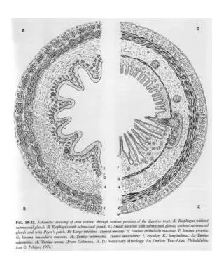
In histologic sections of oral mucosa the basement membrane between the epithelium and connective tissue, which appears as structureless band in H & E stain preparations, stains brightly with periodic acid-Schiff reaction. Ultrastucturally, this region is described as the basal lamina and is highly organized. See Figures
Most components of the basal lamina are synthesized by epithelial cells. • The lamina lucida is made of laminin (glycoprotein). • Laminin cements type IV collagen between the lamina densa and the epithelial cells. • The lamina densa is made of type IV collagen coated with heparan sulphate. • Thick collagen fibres (anchoring fibrils; collagen type VII) attach to the lamina densa to link the basal lamina to the connective tissue.
Fibronectin is sometimes found in the lamina densa and may bind fibroblasts and proteoglycans to it. • The role of the basal lamia: • Provide mechanical adhesion between the epithelium and the connective tissue. • Molecular barrier. • Has a role in response to injury.
Basal Complex Basement membrane: Lamina lucida Lamina densa (Hemidesmosomes)
Lamina propria and submucosa • The lamina propria may be described as a connective tissue of variable thickness that supports the epithelium. • It is divided for descriptive reasons into two parts; papillary and reticular. The lamina propria may attach to the periostium of the alveolar bone, or it may overlay the submucosa, which varies in different regions of the mouth such as the soft palate and floor of the mouth. • The lamina propria contains several different cells: fibroblasts, macrophages, mast cells and inflammatory cells. • The submucosa consists of connective tissue of varying thickness and density. • It attaches the mucous membrane to the underlying structures. • Whether this attachment is loose or firm depends on the character of submucosa. • Glands, blood vessels, nerves, and also adipose tissues are present in this layer. • Submucosa may be present or absent.
Non-keratinized Parakeratinized Orthokeratinized Non-keratinized: Buccal mucosa Labial mucosa Floor of mouth Ventral surface of tongue Posterior 1/3 of dorsal lingual mucosa (including lingual tonsils) Soft palate Alveolar mucosa Junctional epithelium Sulcular epithelium Taste papillae of the tongue (fungiform, foliate and circumvallate) Orthokeratinized or parakeratinized: Hard palate Gingivae (attached gingiva and external side of free gingiva) Anterior 2/3 of dorsal lingual mucosa (filiform papillae) Vermilion zone of the lip Types of Normal Oral Mucosa
Specialized mucosa (dorsal lingual) • Papillae: (filliform, fungiform, circuvallate, foliate) • Taste buds
Functions of oral mucosa • The normal oral mucosa serves several functions. • The major one is protection of deeper tissues of the oral cavity; • others include acting as a sensory organ and • serving as the site of glandular activity and secretion. • However, the human oral mucosa plays no role in regulating body temperature which is not the situation in some animals (such as the dog). • The epithelium affords protection from the mechanical forces of mastication and also acts as a barrier, preventing access of micro-organisms and their toxic products. The mucosa also has a role in immunological defense via the presence of antigen- presenting cells (e.g. Langerhans' cells) and lymphocytes. • The mucosa is well innervated and sensory perception controls the positioning and function of the oral structures important in mastication, swallowing and speech. The mucosa also contains the taste buds and is important in regulating some reflexes such as salivation and gagging.
The mucosa contains many minor salivary glands which help to maintain the moist environment which is necessary for function. • Oral mucosa serves as a barrier but there is a degree of permeability which is of clinical significance (e.g. mouth washes, steroid mouth washes and glyceryl trinitrate in the treatment of angina) • An essential part of the oral environment is the free salivary flow which not only maintains the physiological environment necessary for the maintenance of epithelial integrity but also includes a number of protective, antibacterial components predominantly the IgA. • The microbial flora of the mouth is a further component of the normal healthy oral environment. A change in local or generalised conditions may upset the commensal relationship of the microbial flora of the mouth and the organisms become pathogenic.
Age changes in oral structures Clinically, the oral mucosa of an elderly person often has a smoother and dryer surface than that of a youngster and may be described as atrophic or friable but these changes likely represent the cumulative effects of systemic disease, drug therapy, or both, rather than an intrinsic biologic aging process of the mucosa. The dorsum of the tongue may show a reduction in the number of filiform papillae and a smooth or glossy appearance, such changes being exacerbated by any nutritional deficiency of iron or B complex vitamins. The reduced number of filiform papillae may make the fungiform papillae more prominent, and patients erroneously may consider it to be a disease state. Langerhans' cell become fewer with age, which may contribute to a decline in cell mediated immunity
A striking and common feature in elderly persons is nodular varicose veins on the undersurface of the tongue. • Sebaceous glands (Fordyce's spots) of the lips and cheeks also increase with age, and the minor salivary glands show considerable atrophy with fibrous replacement. • Elderly patients, particularly postmenopausal women, may have symptoms such as dryness of the mouth burning sensation, and abnormal taste. Whether such symptoms reflect systemic disturbances or local tissue changes is not clear. • Recent evidence suggests that stimulated salivary flow rate does not fall purely as a result of age. However, medications or systemic diseases can affect salivary output.