Download
1 / 15
240 likes | 1.16k Views
Oral Mucosa DHY114 – Week 1. Chapter 9. Oral Mucosa. Composed of: stratified squamous epithelium Lining of the oral cavity Hard palate Soft palate Gingiva (attached and free) Buccal and labial mucosa Alveolar mucosa. Classification of Oral Mucosa. Lining mucosa Masticatory mucosa

E N D
Oral MucosaDHY114 – Week 1 Chapter 9
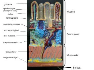
Oral Mucosa • Composed of: stratified squamous epithelium • Lining of the oral cavity • Hard palate • Soft palate • Gingiva (attached and free) • Buccal and labial mucosa • Alveolar mucosa
Classification of Oral Mucosa • Lining mucosa • Masticatory mucosa • Specialized mucosa
Lining of Mucosa • Lining mucosa (stretches, nonkeratinized) • Buccal and labial mucosa • Alveolar mucosa • Floor of the mouth • Ventral surface of the tongue • Soft palate
Masticatory Mucosa • Masticatory Mucosa: (rubbery, keratinized) Gingiva, hard palate, dorsal surface of tongue Torres and Ehrlich Modern Dental Assisting, 7th Ed., Saunders 2002, p. 94, 124.
Specialized Mucosa • Specialized mucosa: Dorsal and lateral aspect of the tongue • Covered with lingual papillae • Fungiform • Circumvallate • Filiform • Foliate
Types of Stratified Squamous Epithelium • Nonkeratinized • Orthokeratinized (keratinized) • Parakeratinized
Nonkeratinization • Nonkeratinization: nuclei present • Examples: Lips, lining mucosa, buccal and labial mucosa, floor of the mouth, ventral surface of the tongue, alveolar mucosa, soft palate, sulcus, col Flattened surface cells Brand and Isselhard. Anatomy of Orofacial Structures. 7th Ed. 2003, p. 294.
Nonkeratinization • Col: (kawl) Depression between the lingual and facial papilla under the contact area. The probe readings Underneath the col may give the first sign of periodontal disease. Wilkins. Clinical Practice of the Dental Hygienist, 8th Ed., 1999, Lippencott, Williams and Wilkins, p. 192.
Keratinization (orthokeratinization) • Keratinized: No nuclei, flat cells • Examples: Masticatory mucosa, which are the hard palate, gingiva, and dorsal surface of tongue Keratinized layer Brand and Isselhard. Anatomy of Orofacial Structures. 7th Ed. 2003, p. 294.
Parakeratinization • Parakeratinization: Shriveled nuclei or no nuclei • Examples: Traumatized hard palate or gingiva Parakeratotic layer Brand and Isselhard. Anatomy of Orofacial Structures. 7th Ed. 2003, p. 294.
Mucocele • Minor salivary gland traumatized, usually on the lip, that causes a blisterlike lesion Ibsen and Phelan. Oral Pathology for the Dental Hygienist, Second Edition, 1996, W. B. Saunders Company, Color Plate 13.
Edema • Swelling: accumulation of fluid in the cells Ibsen and Phelan. Oral Pathology for the Dental Hygienist, Second Edition, 1996, W. B. Saunders Company, Color Plate 13.