Download
1 / 20
200 likes | 286 Views
This study explores the role of Adora2b in stabilizing Per2 to facilitate a metabolic switch critical for myocardial adaptation to ischemia. Using mouse models, differential gene expression during cardiac ischemic preconditioning in Adora2b knockout mice was analyzed, revealing crucial pathways and networks involved. Furthermore, the study delves into the circadian clock network in Adora2b-deficient mice following ischemic preconditioning. Overall, the research sheds light on the molecular mechanisms underlying myocardial adaptation to ischemia.

E N D
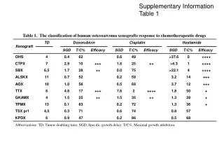
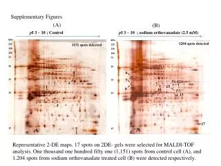
Supplementary Information Figures 1-19 Adora2b-elicited Per2 stabilization promotes a HIF-dependent metabolic switch critical for myocardial adaptation to ischemia Tobias Eckle, Katherine Hartmann, Stephanie Bonney, Susan Reithel, Michel Mittelbronn, Lori A. Walker, Brian D. Lowes, Jun Han, Christoph H. Borchers, Peter M. Buttrick, Douglas J. Kominsky, Sean P. Colgan and Holger K. Eltzschig
Suppl. Figure 1 a Figure S1. Experimental set-up for in situ ischemic preconditioning (IP) and ischemia exposure. (a) In studies of IP-elicited changes in gene expression, preconditioned myocardium was analyzed for transcript or protein levels following indicated time periods of reperfusion.
Suppl. Figure 2 Figure S2. Canonical pathway analysis. Wild-type mice or gene-targeted mice for the Adora2b (Adora2b-/- ) were exposed to ischemic preconditioning (4 cycles consistent of 5 min ischemia followed by 5 min of reperfusion). Following two hours of reperfusion, cardiac tissue from preconditioned myocardium was compared to control tissue without preconditioning. Published online January 14th 2010, NCBI, Gene Expression Omnibus. (http://www.ncbi.nlm.nih.gov/geo/query/acc.cgi?acc=GSE19875). We found a total of 30 differentially regulated genes during cardiac IP comparing WT and Adora2b-/- mice. Canonical pathway analysis was assessed by Ingenuity Software . Only pathways that reached the threshold according to Ingenuity Software analysis are displayed.
Suppl. Figure 3 Figure S3. First of two possible networks based on 30 differentially regulated genes during cardiac IP in wild-type vs. Adora2b-/- mice assessed by Ingenuity Software. Wild-type mice or gene-targeted mice for the Adora2b (Adora2b-/- ) were exposed to ischemic preconditioning (4 cycles consistent of 5 min ischemia followed by 5 min of reperfusion). Following two hours of reperfusion, cardiac tissue from preconditioned myocardium were compared to controls without preconditioning. (red=up regulated genes, green= down regulated genes based on the microarray analysis; comparison wild-type mice over Adora2b-/- mice; drawn connections indicate directly or indirectly interacting genes based on Ingenuity database. published online January 14th 2010, NCBI, Gene Expression Omnibus, http://www.ncbi.nlm.nih.gov/geo/query/acc.cgi?acc=GSE19875).
Suppl. Figure 4 Figure S4. Second of two possible networks based on 30 differentially regulated genes during cardiac IP in wild-type vs. Adora2b-/- mice assessed by Ingenuity Software. Wild-type mice or gene-targeted mice for the Adora2b (Adora2b-/- ) were exposed to ischemic preconditioning (4 cycles consistent of 5min ischemia followed by 5 min of reperfusion). Following two hours of reperfusion, cardiac tissue from preconditioned myocardium were compared to controls without preconditioning. (red=up regulated genes, green= down regulated genes based on the microarray data; regulation means wild-type mice over Adora2b-/- mice; drawn connections indicate directly or indirectly interacting genes based on Ingenuity database published online January 14th 2010, NCBI, Gene Expression Omnibus, http://www.ncbi.nlm.nih.gov/geo/query/acc.cgi?acc=GSE19875).
Suppl. Figure 5 a Wildtype Adora2b-/- Clock Actb IP C C IP Fold change 1.1 Fold change 0.8 Micro Array IP vs C b Wildtype Adora2b-/- Cry1 Actb C IP C IP Fold change 1.0 Fold change 0.9 Micro Array IP vs C Adora2b-/- c Wildtype Timeless Actb C IP C IP Fold change 1.3 Micro Array IP vs C Fold change 0.9 d Wildtype Adora2b-/- Prkaa1 Actb C C IP IP Fold change 1.2 Fold change 0.9 Micro Array IP vs C Figure S5. Analysis of the circadian clock network in Adora2b-/- mice following cardiac IP. Adora2b-/-mice or littermate controls matched in age, weight and gender were subjected to in situ IP treatment consistent of 4 cycles of IP (5 minutes of ischemia, 5 minutes of reperfusion). Cardiac preconditioned tissue was shock-frozen and analyzed for protein levels. (a-d) Western blot of Clock (Clock), Cryptochrome 1 (Cry1), Timeless (Timeless) and AMP-activated protein kinase, alpha 1 (Prkaa1). Note: Microarray data revealed no changes for Clock, Cry1 or Prkaa1 in contrast to Western Blot analysis.
Suppl. Figure 6 a Figure S6. Experimental set-up for in situ ischemic preconditioning (IP) and ischemia exposure. (a) In vivo model of myocardial ischemic preconditioning and subsequent determination of myocardial injury. Following induction of anesthesia (A) and thoracotomy (T), mice were exposed to 60 minutes of myocardial ischemia with or without previous IP pretreatment. After 120 min of reperfusion, the area at risk was determined by retrograde injection of Evan’s blue, cardiac tissues were harvested and infarct staining was performed using 2,3,5-triphenyltetrazolium chloride (TTC). Myocardial injury was assessed by measuring infarct sizes determined as percentage of the infarcted area from the area at risk or by measurements of plasma troponin I levels.
Suppl. Figure 7 a Wildtype Adora2b-/- Per1 Actb C C IP IP Fold change 0.8 Fold change 2.0* Micro Array IP vs C b C-Adora2b-/- IP-WT C-WT IP-Adora2b-/- Per1 IHC Per1-/- Hif1a c d e Actb C IP Figure S7. Per1 in myocardial ischemia. Adora2b−/− or littermate control mice matched in age, gender and weight were subjected to in situ preconditioning with 4 cycles of IP (5 minutes of ischemia, 5 minutes of reperfusion). (a,b) Per1 protein levels determined by Western blot (a) or immunohistochemistry(b) following IP-treatment without reperfusion. One representative image of three is displayed. * significant differential regulation based on microarray data (c) Per1-/- were exposed to 60 min of in situ myocardial ischemia followed by 2h of reperfusion, infarct sizes were measured by double staining with Evan’s blue and triphenyltetrazolium chloride or Troponin I serum levels (mean±SD; n=6, scale bare 50 mm) (d) Glycogen content of Per1-/- hearts IP (mean±SD; n=6). (e) Western blot for Hif1a after cardiac IP treatment. One representative blot of three is displayed.
Suppl. Figure 8 a Figure S8. In vitro model of hypoxic preconditioning. For in vitro preconditioning with hypoxia, cardiomyocytes were exposed to 3 cycles of 45 minutes hypoxia (1%) followed by 20 minutes of normoxia (21%). Following 180 min of normoxia exposure, cells were harvested and analyzed for transcript levels by real-time RT-PCR or for protein levels by Western blot analysis, respectively.
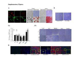
Suppl. Figure 9 a IHD Control PER2 ACTB 1 2 3 4 1 2 3 4 Patient No. Densitometry 1 1 0.8 1.7 3.2 2.26 3.56 1.56 b Control IHD PER2 ACTB Patient No. 5 6 7 5 6 7 Densitometry 1 1.9 2.1 3.57 2.8 3.4 c Control IHD PER2 ACTB Patient No. 8 9 10 8 9 10 Densitometry 1 0.9 1.2 3.6 2.4 2.2 Figure S9. Per2 Western blot analysis obtained from cardiac biopsies of patients suffering from severe ischemic heart disease (IHD) or healthy controls (cardiac donations). For statistical analysis and overview, see Figure 1h; patient characteristics are given in Table S1.
Suppl. Figure 10 a b c d HMEC SC HMEC ADORA2BKD PER2 ACTB BAY 60-6583 [ZT] 0 6 12 0 6 12 Figure S10. Validation of human microvasuclar endothelial cells (HMEC-1) as in vitro model for PER2 regulation. (a) Transcript levels of adenosine receptors in HMEC-1 determined by real time RT PCR (mean±SD, n=3). (b) ELISA for cAMP in HMEC-1 after ADORA2B agonist (10 mM, BAY 60-6583) or forskolin (30 mM) treatment (mean±SD, n=3). (c) ELISA for Phospho-CREB in wildtype HMEC-1 (HMEC SC, treated with scrambled siRNA) or ADORA2B knockdown HMEC-1 (HMEC ADORA2B KD, treated with ADORA2B siRNA) after ADORA2B agonist (10 mM, BAY 60-6583) treatment (mean±SD, n=3). (d) Synchronized HMEC SC or HMEC ADORA2B KD cells were treated with vehicle, or ADORA2B agonist BAY 60-6583 and blotted after indicated time periods; one of three representative experiments is displayed.
Suppl. Figure 11 a b c d Figure S11. PER2 promoter analysis. (a) Chromosomal localization of PER2. (b) PER2 promoter with putative CREB binding sites (yellow) and truncations (green/bold) used in promoter assays. (c) Nucleotide sequence alignments of conserved regions shared by human and mouse Per2 promoter. H and M represent human and mouse sequences, respectively. The bold , underlined letters represent a conserved CREB binding site between human and mouse, which was identified to be functional (Fig. 1i). Promoter analysis was performed using Genomatix Software (http://www.genomatix.de) and TESS (http://www.cbil.upenn.edu/cgi-bin/tess). Alignment was performed using BLAST (NCBI). (d) Schematic of plasmids expressing sequence corresponding to full length PER2 promoter (FLPER2) or indicated truncations: -88, -216, -357,-461 with putative CREB binding sites (red = CREB binding site that significantly increased luciferase activity over baseline activity upon ADORA2B activation, see Fig. 1j).
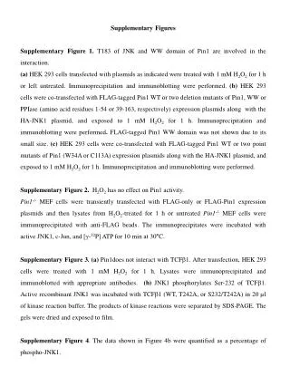
Suppl. Figure 12 b a CXM+FSK Vehicle CXM FSK PER2 ACTB ZT6 d c Vehicle BAY 60-6583 CULNEDD8 ACTB 6 24 ZT 0 12 6 24 12 csiR e siCSN5 f - - - BAY 60-6583 + + + - - - - siCSN5 + + - - - - csiR + + Vehicle - - - - - + CSN5 ACTB 6 ZT 0 Figure S12. In vitro studies on ADORA2B dependent CUL deneddylation as molecular mechanism for PER2 stabilization. (a)Validation of actinomycin (ACT) and cycloheximide (CXM) treatment for studies on the influence of transcriptional, translational or post-translational mechanisms on Period 2 protein levels (Fig. 2b,c). (a,left) Inhibition of transcription using ACT. # significant inhibition of Per2 transcription; * significant increase of Per2 transcript due to pharmacological treatment (forskolin, FSK). (b, right) Inhibition of translation using CXM. (Fig. 2b,c).(b) Neddylated cullin enhances ubiquitin ligase (SCF) activity leading to proteasomal degradation. The COP9 signalosome or signaling via ADOAR2B leads to cullin deneddylation. (c,d) Synchronized HMEC-1 treated with vehicle or ADORA2B agonist BAY 60-6583 (10mM) were blotted after indicated time periods; one of three representative experiments is displayed, and quantified for neddylated CULLIN using a specific Nedd8 antibody (*p<0.05, n=3). (e, f) siRNA knockdown of the COP9 signalosome subunit CSN5. RT PCR or western blot for CSN5 from HMEC-1 after siRNA treatment. Note: siRNA knockdown revealed a 98 % reduction of CSN5 transcript or protein, respectively.
Suppl. Figure 13 a c b d e wildtype Per2 -/- f Gys1 Actb Baseline WT Per2-/- Figure S13. Baseline characteristics of Per2-/-mice. (a) Cardiac characterization of Per2-/- mice. Per2 transcript levels were measured in cardiac tissues by real-time RT-PCR relative to housekeeping gene β-actin. (WT: wildtype; HZ - heterozygote, KO - homozygous deletion of Per2, n=3). (b,c) Hearts from wildtype and Per2-/- mice were analyzed for glycogen (b) or long chain fatty acids (c) using an enzymatic ELISA KIT from Biovision (mean±SD; n=3). (d, e) Baseline protein levels of glycogen synthase 1(Gys1,d) or carnitine-palmitoyltransferase I (Cpt1,e). One representative blot of three is displayed. (f) Echocardiographic analysis of hearts at baseline comparing wild-type and Per2-/- mice (EF= ejection fraction [%], LVEDV=left ventricular end diastolic volume, mean±SD; n=3). wildtype Per2-/- Cpt1 Actb Baseline
WT Per2-/- Suppl. Figure 14 d a e b f c j g h i k l Figure S14. Consequences of ischemic preconditioning (IP) on glucose metabolism in wild-type or Per2-/- mice. Per2-/- mice or littermate controls matched in age, weight and gender were subjected to IP, ischemia alone or ischemia with IP (IP; 4 cycles of 5 min ischemia followed by 5 min of reperfusion). (a-j) Cardiac transcript levels of glycolytic enzymes determined by real-time RT-PCR relative to Actb and expressed as fold induction relative to sham-operated controls (mean±SD n=3), hexokinase 4 (Hk4), phosphofructokinase-M (Pfkm), glyceraldehyde 3-phosphate dehydrogenase (Gapdh), phosphoglycerate kinase 1 (Pgk1), pyruvate kinase (Pk), pyruvate dehydrogenase kinase isozyme 1, 2 and 4 (Pdk1, Pdk2, Pdk4), lactate dehydrogenase a and b (Ldha, Ldhb). (k, l) Pyruvate kinase and lactate dehydrogenase enzyme activity (mean±SD n=3).
WT Per2-/- Suppl. Figure 15 a b c e d Figure S15. Consequences of Period 2 deficiency on cardiac metabolism during myocardial ischemia or during reperfusion with or without IP pretreatment. (a-e) Per2-/- mice or littermate controls matched in age, weight and gender were exposed to 60 min of in situ myocardial ischemia and 60 minutes of reperfusion with or without ischemic preconditioning (IP; 4 cycles of 5 min ischemia followed by 5 min of reperfusion) prior to myocardial ischemia. C13-glucose for the studies during the ischemic period were administered 30 minutes prior to the onset of ischemia while for studies during the reperfusion phase, we applied the tracers at the onset of reperfusion. Both times the tracer was applied via intravascular injection into a catheter placed into the carotid artery. Isotopically labeled 13C-glucose was purchased from Cambridge Isotope Labs . All UPLC-MS data were acquired with a Waters Acquity UPLC system coupled to a Water Synapt HDMS quadrupole time-of-flight mass spectrometer. Metabolites were measured from the area at risk. (a) 13C-glucose (b) 13C-fructose-1,6-bisphosphate. (c) 13C-lactate. (d) Glucose oxidation (TCA cycle flux rates determined by the ratio of 13C3-glutamate and total creatine). (e) Glycogen content assessed by Glycogen Assay Kit. Per2-/- mice exhibited enhanced glycogen content at baseline, however did not recover during reperfusion (mean±SD; n=3).
Suppl. Figure 16 b a c d Figure S16. Hif1a regulation in Per2-/- mice. Analysis of cardiac transcript levels from wild-type or Per2-/-hearts during a 24 h period for Hif1a isoforms Hif 1.1 and 1.2, Pdk1 and Ldh1. (a) Hif 1.1 (b) Hif 1.2. (c) Pdk1 (pyruvate dehydrogenase kinase isozyme 1). (d) Ldha (lactate dehydrogenase a). Real-time RT-PCR relative to Actb and expressed as fold change of transcript relative to wild-type controls at ZT0 (mean±SD, n=3, #,*p<0.05 over control).
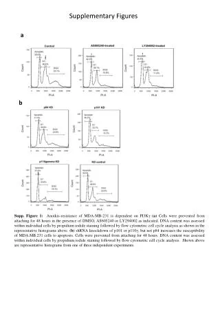
Suppl. Figure 17 a Cre+ DMOG Cre+ control CardiacHif1a-/-DMOG b Cre+ myocytes CardiacHif1a-/- myocytes Hif1a Actb [4h] Hx Hx Nx Nx Nx 21 % O2 Hx 1 % O2 Figure S17. Characterization of cardiac specific Hif1a-/-mice. (a) Comparison of immunoreactivity for Hif1a in untreated controls (Cre+) versus DMOG treated cardiac tissue from controls (Cre+) or from animals with a Hif1aloxp/loxpMyosin‐Cre+ background (cardiacHif1a-/- , magnification x 20, one of three representative images is displayed). Note: Hearts from animals with a HIF1aloxp/loxpMyosin‐Cre background (cardiacHif1a-/-) under TMX display absent to very low sarcoplasmatic Hif1a expression compared to strong sarcoplasmic and nuclear Hif1a expression seen in Cre+ animals with DMOG; scale bar 50 mm. (b) Adult Myocytes were isolated from controls (Cre+) or from animals with a Hif1aloxp/loxp Myosin‐Cre+ background (cardiacHif1a-/- ) and exposed to ambient hypoxia [1%, 4h]. One representative blot of three is displayed.
Suppl. Figure 18 b a d c Figure S18. Hif1a regulation in Adora2b-/-. Analysis of cardiac Hif1a isoforms Hif 1.1 (a) and Hif 1.2 (b), Pdk1 (pyruvate dehydrogenase kinase isozyme 1, c) and Ldha (lactate dehydrogenase a, d) transcript levels from wild-type or Adora2b-/-hearts during a 24 h time period. Real-time RT-PCR relative to Actb and expressed as fold change of transcript relative to wild-type controls at ZT0 (mean±SD, n=3, #, *p<0.05 over control).
Suppl. Figure 19 a Per2 Actin 0 6 12 ZT b c WT Room light WT Day light Per2-/- Day light Figure S19. Light-induced stabilization of cardiac Per2 provides potent protection from myocardial ischemia. (a) Naturally occurring stabilization of cardiac Per2 where ZT 0 is the start of the subjective day. (b) Per2-/- mice or littermate controls matched in age, gender and weight underwent light therapy as described above over indicated time periods, followed by exposure to in situ myocardial ischemia (60 min) followed by 2h of reperfusion. Myocardial injury was assessed by measurement of infarct size or troponin I plasma levels (Fig. 6i; n=6 mice per experimental group, mean±SD); scale bar 500 mm (c) Wild-type mice were subjected to in situ myocardial ischemia (60 min) followed by 2h of reperfusion over a 24 h time period (mean±SD). Myocardial injury was assessed by measurement of infarct size or troponin I plasma levels (Fig. 6k; n=8 mice per experimental group, mean±SD).