Download
1 / 33
330 likes | 338 Views
Learn about the functions, organization, and structure of the nervous system, including neurons and neuroglia. Understand the central and peripheral nervous systems and the autonomic nervous system.

E N D
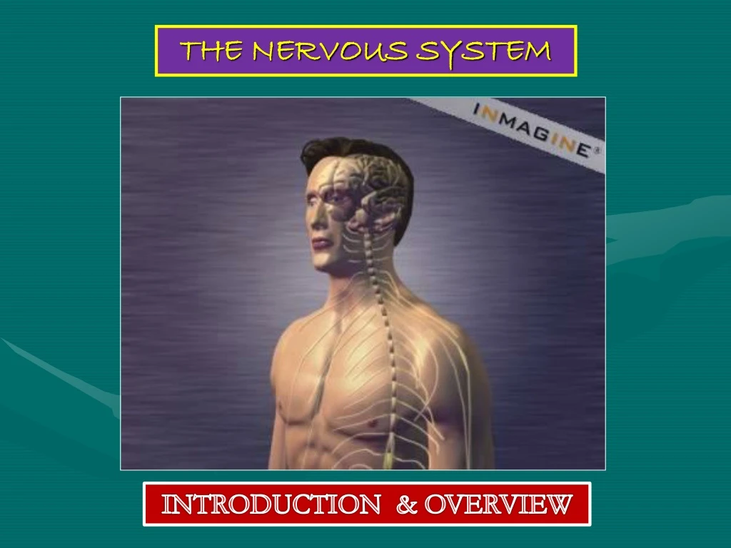
THE NERVOUS SYSTEM INTRODUCTION & OVERVIEW
FUNCTIONS • collection of sensory input • integration • motor output The function of the nervous system is to detect changes in the: External or Internal environments So, it bring about appropriate responses in Muscles, Organs and Glands.
ORGANIZATION STRUCTURAL • CNS • PNS FUNCTIONAL • Sensory division (Afferent) • Motor division (Efferent) • autonomic • somatic
Neurons It is the basic structural (anatomical) , functional and embryological unit of the nervous system. The human nervous system is estimated to contain about 1010. What is neurone? Prof. Saeed Makarem
The functions of the neuron is to receive and integrate incoming information from sensory receptors or from other neurons and to transmit information to other neurons or effector organs. Prof. Saeed Makarem
Information is passed between neurons at specialized regions called synapses In the synapses the membranes of adjacent cells are in close apposition (contiguity not continuity). Prof. Saeed Makarem
There is wide diversity in the shape and size of neurons in different parts of the nervous system. • But all share certain common characteristics. • There is a single cell body from which a variable number of branching processes emerge. Prof. Saeed Makarem
Most of these processes are receptive in function. They are known as Dendrites. Prof. Saeed Makarem
The dendrites possess: A variable number of shot processes. They form the receptive element of the neurone. Prof. Saeed Makarem
One of the processes leaving the cell body is called the axonwhich carries information away from the cell body. • Axons are highly variable in length and may divide into several branches or collaterals through which information can be distributed to a number of different destinations simultaneously. • At the end of the axon, specializations called terminal buttons occur. • Here information is transferred to the dendrites of other neurones. Prof. Saeed Makarem
Transmission of information between neurons almost always occurs bychemicalrather than electrical means. • Action potential causes release of specific chemical that are stored in synaptic vesicles in the presynaptic ending. • These chemicals are known as neurotransmitters and diffuse across the narrow gap between pre- and postsynaptic membranes to bind to receptorson the postsynaptic cell. Prof. Saeed Makarem
Neuroglia • Neuroglia, or gliacells constitute the other major cellular component of the nervous system. • It is a specialized connective tissue for the nervous system. • Unlike neurones, neuroglia do not have a direct role in information processing but they are essential for the normal functioning of nerve cells. Prof. Saeed Makarem
Three main types of neuroglial cell are recognized: • Oligodendrocytes(oligodendroglia) they form the myelin sheaththat surrounds many neuronal axons, which increase the rate of conduction. Prof. Saeed Makarem
2- Microglia have a phagocytic role in response to nervous system damage. Prof. Saeed Makarem
Astrocytesare thought to form a selectively permeable barrier between the circulatory system and the neurons of the brain and spinal cord. • This is known as the 'blood-brain barrier' and has a protective function. Prof. Saeed Makarem
The nervous system is divided into: • 1- Central nervous system (CNS). • 2- Peripheral nervous system (PNS). Prof. Saeed Makarem
The central nervous system consists of the brain and the spinal cord,lying within the protection of the cranium and vertebral column, respectively. • This is the most complex part of the nervous system. • It contains the majority of nerve cell bodies and synaptic connections. Prof. Saeed Makarem
The peripheral nervous system constitutes the link between the CNS & structures in the periphery of the body. • It receives sensory information from the body. • It sends controlling impulses in response to these information. • The peripheral nervous system consists: • 1- Cranial nerves • 2- Spinal nerves. Prof. Saeed Makarem
Spinal nerves supplying the upper or lower limbs form the brachialor lumbar plexus. • Nerve cell bodies that are aggregated within the CNS are called GANGLIA Prof. Saeed Makarem
Autonomic Nervous System • Neurones that detect changes and control the activity of, the viscera are collectively referred to as the autonomic nervous system. • Its components are present in both the central and peripheral nervous systems. Prof. Saeed Makarem
SYMPATHETIC & PARASYMPATHETIC SYSTEMS • The autonomic nervous system is divided into two anatomically and functionally distinct parts: • Sympathetic: Or • Thoracolumbar outflow • Parasympathetic: Or • Craniosacral outflow.
Sympathetic and parasympathetic divisions are generally have antagonistic effects on the structures that they innervate. Prof. Saeed Makarem
The autonomic nervous system innervates: • Smooth muscle, • Cardiac muscle, • Secretory glands. • It is an important part of the homeostatic mechanisms that control the internal environment of the body. Prof. Saeed Makarem
Afferent, Efferent & Interneuron • Nerve cells that carry information from peripheral receptors to the CNS are referred to as afferent neurones • Efferentneurones carry impulses away from the CNS • If they innervate skeletal muscle to cause movement they are also referred to as motor neurons. • The vast majority of neurones, however, lies entirely within the CNS and are usually called interneuron
PARTS OF THE BRAIN • Cerebral hemispheres • Diencephalon • Cerebellum • Brain stem
CEREBRAL HEMISPHERES • The largest part of the brain • They have elevations, called gyri • Gyri are separated by depressions called sulci • Each hemisphere is divided into 4lobes • Lobes are separated by deeper grooves called fissures or sulci. PARIETAL FRONTAL TEMPORAL OCCIPITAL
TISSUE OF THE CEREBRAL HEMISPHERES • The outer layer is the gray matter or cortex • Deeper is located the white matter, composed of bundles of nerve fibers, carrying impulses to and from the cortex • Basal nucleiare made from gray matter and are located deep within the white matter • They help the motor cortex in the regulation of voluntary motor activities Basal nuclei
DIENCEPHALON The diencephalon is located between the 2 hemispheres and is linked to them and to the brainstem. The major structures of the diencephalon are thethalamus, hypothalamus and Epithalamus.
BRAIN STEM The brainstem has three parts: midbrain, Pons and medulla oblongata.
CEREBELLUM Cerebellum has 2 cerebellar hemispheres and a convoluted surface. It has an outer cortex of gray matter and an inner region of white matter. It provides precise coordination for body movements and helps maintain equilibrium.
BRAIN VENTRICLES • Brain is bathed by the cerebrospinal fluid (CSF) • Inside the brain, there are spaces (ventricles) filled with CSF • There are 4 ventricles • 2lateral ventricles: are in the hemispheres • 3rd ventricle: in the diencephalon • 4th ventricle: between the Pons & the cerebellum • Cerebral aqueduct: connects the 3rd to the 4th ventricle
CSF is constantly produced by the choroid plexuses inside each ventricle. CEREBROSPINAL FLUID Inside the brain, CSF flows from the lateral ventricles to the 3rd and 4thventricles. Most of the CSF drains from the 4th ventricle in the subarachnoid space around the brain and returns to the dural sinuses through the arachnoids villi. From the 4th ventricle, part of the CSF flows down in the central canal of the spinal cord.


