GLUCONEOGENESIS
GLUCONEOGENESIS. The synthesis of glucose from noncarbohydrate precursors Mainly in the liver. IMPORTANT PRINCIPLES OF BIOSYNTHESIS. The synthesis pathway is usually different from the degradation pathway. The two opposing pathways may share many reversible reactions.

GLUCONEOGENESIS
E N D
Presentation Transcript
GLUCONEOGENESIS The synthesis of glucose from noncarbohydrate precursors Mainly in the liver
IMPORTANT PRINCIPLES OF BIOSYNTHESIS • The synthesis pathway is usually different from the degradation pathway. • The two opposing pathways may share many reversible reactions. • There is always at least one unique enzymatic step to each pathway. BECAUSE If this wasn’t the case, then the flow of carbon through the two pathways would be dictated by the mass action and not the cellular changing needs for energy, precursors or macromolecules.
IMPORTANT PRINCIPLES OF BIOSYNTHESIS • Corresponding anabolic and catabolic pathways are controlled by different regulatory enzymes in a reciprocal manner. • If one pathway is stimulated, then the opposite is inhibited. • Biosynthetic pathways are usually regulated at their initial steps. BECAUSE To prevent wasting precursors to make unneeded intermediates.
IMPORTANT PRINCIPLES OF BIOSYNTHESIS • The energy-requiring biosynthetic processes are coupled to the energy-yielding hydrolysis of ATP • The overall process is essentially irreversible in vivo. • The total amount of energy from ATP (and NAD(P)H) is larger than the minimum energy needed to convert the precursor into the biosynthetic product. THUS The resulting large negative free energy for the overall process will assure that it will take place even when the concentrations of the precursors are relatively low.
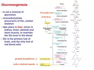
Triglycerols Fatty acids glycerol Dietary & muscle proteins Amino acids Noncarbohydrate precursors of glucose:
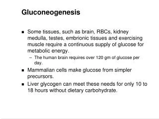
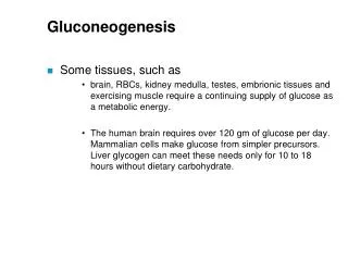
Main sites of gluconeogenesis: • Major site: Liver. • Minor site: Kidney. • Very little: • Brain. • Muscle (skeletal and heart). gluconeogenesis in the liver and kidney helps to maintain the glucose level in the blood so that brain and muscle can extract sufficient glucose from it to meet their metabolic demands.
Gluconeogenesis Is Not a Reversal of Glycolysis: • Seven steps are shared by glycolysis and gluconeogenesis. • However, three essentially irreversible steps in glycolysis shift the equilibrium far in the side of glycolysis. • Most of the decrease in free energy in glycolysis takes place in these steps.
In gluconeogenesis the three reactions are bypassed by a set of separate enzymes. • Phosphoenolpyruvate is formed from pyruvate: • Fructose 6-phosphate is formed from fructose 1,6-bisphosphate: • Glucose is formed by hydrolysis of glucose 6-phosphate:
A B PYRUVATEPHOSPHOENOLPYROVATE
PYROVATE CARBOXYLASE Mitochondrial enzyme
1 2 3 CARBOXYLATION OF PYROVATE: HCO3-, the aqueous form of CO2 is activated to carboxyphosphate. it is subsequently activated by binding to the N-1 atom of the biotin ring to form the carboxybiotin-enzyme intermediate. The DG°´ for its cleavage is -20 kJ mol-1. The activated carboxyl group is then transferred from carboxybiotin to pyruvate to form oxaloacetate.
The long, flexible link between biotin and the enzyme enables this prosthetic group to rotate from one active site of the enzyme (the ATP-bicarbonate site) to the other (the pyruvate site). • To be carboxylated, biotin needs the enzyme to be allosterically activated by Acetyl CoA.
DECARBOXYLATION AND PHOSPHORYLATION OF OXALOACETATE: • Oxaloacetate, is reduced to malate inside the mitochondrion for transport to the cytosol. • The reduction is accomplished by an NADH-linkedmalate dehydrogenase. • When malate has been transported across the mitochondrial membrane, it is reoxidized to oxaloacetate by an NAD+-linkedmalate dehydrogenase in the cytosol.
Oxaloacetate is simultaneously decarboxylated and phosphorylated by phosphoenolpyruvate carboxykinase in the cytosol. • The CO2 that was added to pyruvate by pyruvate carboxylase comes off in this step. • The formation of the unstable enol is driven by decarboxylation, and trapped by the addition of a phosphate to carbon 2 from GTP.
NADH + H+ H++ NADH Cytosolic PEP CARBOXYKINASE Oxaloacetate PEP Cytosolic Malate DEHYDROGENASE CO2 NAD+ Malate Malate NAD+ Oxaloacetate Pyruvate CARBOXYLASE CO2 Pyruvate Pyruvate
This pathway predominates when pyruvate or alanine is the glucogenic precursor. • The carboxylation-decarboxylation represents a way of activating pyruvate. • the decarboxylation of Oxaloacetate facilitates PEP formation. • The cytosolic NADH is consumed by other gluconeogenesis reactions, and has to be regenerated in order to proceed with the process. • This is accomplished by transporting Malate outside the mitochondria.
NADH + H+ Cytosolic PEP CARBOXYKINASE Oxaloacetate PEP Cytosolic Malate DEHYDROGENASE NADH + H+ CO2 NAD+ Malate PEP Malate Mitochondrial PEP CARBOXYKINASE NAD+ CO2 H++ NADH Oxaloacetate Oxaloacetate Pyruvate CARBOXYLASE Pyruvate CARBOXYLASE CO2 CO2 Pyruvate Pyruvate Pyruvate Pyruvate NAD+ Lactate DEHYDROGENASE Lactate
This pathway predominates when lactate is the precursor. • The conversion of lactate to pyruvate in the hepatocyte cytosole yields NADH. • Thus no Malate transport is needed any more for this purpose. • The mitochondrial and cytosolic PEP CARBOXYKINASE enzymes are encoded by separate nuclear genes. (two different enzymes catalyzing the same reaction in different localizations)
FRUCTOSE 1,6-BISPHOSPHATEFRUCTOSE 6-PHOSPHATE • The enzyme responsible for this step is fructose 1,6-bisphosphatase. • Like its glycolytic counterpart, it is an allosteric enzyme that participates in the regulation of gluconeogenesis.
GLUCOSE 6-PHOSPHATEGLUCOSE • This hydrolytic reaction is catalyzed by glucose-6-phosphatase in the Endoplasmic reticulum of the hepatocytes.. • In most tissues, free glucose is not generated; • the glucose 6-phosphate is processed in some other fashion, notably to form glycogen. • Unlike free glucose, glucose 6-phosphate cannot diffuse out of the cell.
To keep glucose inside the cell, the generation of free glucose is controlled in two ways: • The enzyme responsible for the conversion of glucose 6-phosphate into glucose, glucose 6-phosphatase, is regulated. • The enzyme is present only in tissues whose metabolic duty is to maintain blood-glucose homeostasis tissues that release glucose into the blood (the liver and to a lesser extent the kidney).
Several endoplasmic reticulum (ER) proteins play a role in the generation of glucose from glucose 6-phosphate. • T1 transports glucose 6-phosphate into the lumen of the ER. • T2 and T3 transport Pi and glucose, respectively, back into the cytosol. • Glucose 6-phosphatase is stabilized by a Ca2+-binding protein (SP).
The rate of conversion of glucose into pyruvate is regulated to meet two major cellular needs: • The production of ATP, generated by the degradation of glucose. • The provision of building blocks for synthetic reactions, such as the formation of fatty acids.
It is all in the enzymes • Enzymes can enhance the rates of metabolic (or other) reactions by many orders of magnitude. • A rate enhancement of 1017 means that what would occur in 1 second with an enzyme’s help, would otherwise require 31,710,000,000 years to take place. • So essentially without enzymes such reactions don’t take place. • Thus, regulation of enzymatic activity is in a sense, regulation of metabolism, or any other cellular process.
Regulation and control of enzyme activity • Substrate level control. • Allosteric effectors • Covalent modification • Enzyme concentration: increased synthesis • Enzyme concentration: generation of active enzyme by processing • Substrate cycles
Substrate level control • Since most often [S] > Km, the change in substrate concentration does not change the reaction rate appreciably. • Thus, controlling a metabolic flux is not normally achieved by varying substrate concentrations.
Allosteric effectors • Noncovalently bind and regulate the enzyme. • The effector may be stimulatory or inhibitory. • The substrate and effector usually occupy different specific binding sites.
Allosteric enzymes kinetics: • Sigmoid kinetic behavior is seen. • K0.5 represents the substrate concentration at which the enzyme velocity is half Vmax. • (-) and (+) respectively indicate inhibitory and stimulatory effectors.
Enzyme concentration: generation of active enzyme by processing
Regulation of the flux through multistep pathways occurs at steps that are enzyme limited: • RATE-LIMITING STEP: the rate of at least one reaction in every metabolic pathway depends on the activity of the enzyme (ENZYME-LIMITED), and is not limited to the substrate availability.
Any enzyme that catalyzes the 1st step in a pathway is a potential control point since it shows “commitment” to the pathway. • Phosphofructokinase is the obvious point in glycolysis. • Any enzyme that is working slowly (small Vmax) is obviously a bottle-neck in the reaction. • Therefore activation of a slow enzyme can increase the flux of the entire pathway. • In heart muscle glycolysis the slowest enzymes are: • Hexokinase. • Phosphofructokinase. • Aldolase. • Enolase.
Irreversible reactions in glycolysis (rate-limiting) are potential sites of control. • Hexokinase • Phosphofructokinase • pyruvate kinase. • Their activities are regulated by: • the reversible binding of allosteric effectors • by covalent modification. • the amounts of these important enzymes are varied by the regulation of transcription to meet changing metabolic needs.
Gluconeogenesis and Glycolysis Are Reciprocally Regulated • The amounts and activities of the distinctive enzymes of each pathway are controlled so that both pathways are not highly active at the same time. • The interconversion of fructose 6-phosphate and fructose 1,6-bisphosphate is stringently controlled. • Phosphofructokinase and fructose 1,6-bisphosphatase are reciprocally controlled by fructose 2,6-bisphosphate in the liver
PHOSPHOFRUCTOKINASE: The most important control element in the mammalian glycolytic pathway. • PFK in the liver is a tetramer of 4 identical subunits. • The allosteric effectors binding site is distinct from the catalytic site.
ATP allosterically inhibit the enzyme: • High concentrations of ATP converts the hyperbolic binding curve of F6-P to sigmoidal one. • AMP reverses the inhibitory effect of ATP • The activity of the enzyme increases when the ATP/AMP ratio is lowered glycolysis is stimulated as the energy charge falls