Gluconeogenesis
Gluconeogenesis. NCVI T.A. Overview. Some tissues, such as the brain, red blood cells, kidney medulla, lens and cornea of the eye, testes, and exercising muscle, require a continuous supply of glucose as a metabolic fuel.

Gluconeogenesis
E N D
Presentation Transcript
Gluconeogenesis NCVI T.A
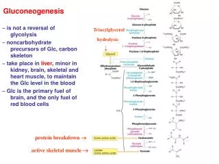
Overview • Some tissues, such as the brain, red blood cells, kidney medulla, lens and cornea of the eye, testes, and exercising muscle, require a continuous supply of glucose as a metabolic fuel. • Liver glycogen, an essential postprandial source of glucose, can meet these needs for only ten to eighteen hours in the absence of dietary intake of carbohydrate. • During a prolonged fast, however, hepatic glycogen stores are depleted, and glucose is formed from precursors such as lactate, pyruvate, glycerol (derived from the backbone of triacylglycerols), and α-ketoacids (derived from the catabolism of glucogenic amino acids). • The formation of glucose does not occur by a simple reversal of glycolysis, because the overall equilibrium of glycolysis strongly favors pyruvate formation. • Instead, glucose is synthesized by a special pathway, gluconeogenesis, that requires both mitochondrial and cytosolic enzymes. • During an overnight fast, approximately ninety percent of gluconeogenesis occurs in the liver, with the kidneys providing ten percent of the newly synthesized glucose molecules. • However, during prolonged fasting, the kidneys become major glucose-producing organs, contributing an estimated forty 40% of the total glucose production. • Figure 10.1 shows the relationship of gluconeogenesis to other important reactions of intermediary metabolism
Substrates for Gluconeogensis • Gluconeogenic precursors are molecules that can be used to produce a net synthesis of glucose. • They include the intermediates of glycolysis & the TCA. • Glycerol, lactate & the α-keto acid obtained from the determination of glucogenic amino acids are the most important gluconeogensis precursors.
glycerol • Glycerol is released during the hydrolysis of triacylglycerols in adipose-tissue, and is delivered by the blood to the liver. • glycerol is phosphorylated by glycerol kinase to glycerol phosphate, which is oxidized by glycerol phosphate dehydrogenase to dihydroxyacetone phosphate—an intermediate of glycolysis. • Note: Adipocytes cannot phosphorylate glycerol because they essentially lack glycerol kinase.
Lactate • Lactate is released into the blood by exercising skeletal muscle, and by cells that lack mitochondria, such as red blood cells. In the Cori cycle, blood borne glucose is converted by exercising muscle to lactate, which diffuses into the blood. • This lactate is taken up by the liver and reconverted to glucose, which is released back into the circulation (Figure 10.2).
Amino Acid • Amino acids derived from hydrolysis of tissue proteins are the major sources of glucose during a fast. α-Ketoacids, such as oxaloacetate (OAA) and α -ketoglutarate, are derived from the metabolism of glucogenic amino acids. • These α -ketoacids can enter the citric acid cycle and form oxaloacetate—a direct precursor of phosphoenolpyruvate (PEP). • Note: Acetyl coenzyme A (CoA) and compounds that give rise to acetyl CoA (for example, acetoacetate and amino acids such as lysine and leucine) cannot give rise to a net synthesis of glucose. • This is due to the irreversible nature of the pyruvate dehydrogenase reaction, which converts pyruvate to acetyl CoA. These compounds give rise instead to ketone bodies and are therefore termed ketogenic.
Reaction unique to gluconeogensis • Seven glycolytic reactions are reversible and are used in the synthesis of glucose from lactate or pyruvate. • However, three of the reactions are irreversible and must be circumvented by four alternate reactions that energetically favor the synthesis of glucose. • These reactions, unique to gluconeogenesis, are described below.
a. Caboxylation of pyruvate • The first "roadblock" to overcome in the synthesis of glucose from pyruvate is the irreversible conversion in glycolysis of PEP to pyruvate by pyruvate kinase. • In gluconeogenesis, pyruvate is first car-boxylated by pyruvate carboxylase to OAA, which is then converter to PEP by the action of PEP-carboxykinase (Figure 10.3).
1. Biotin is a coenzyme • Pyruvate carboxylase requires biotin covalently bound to the ε-amino group of a lysine residue in the enzyme {see Figure 10.3). • This covalently bound form of biotin is called biocytin. Hydrolysis of ATP drives the formation of an enzyme-biotin-CO2 intermediate. • This high-energy complex subsequently carboxylates pyruvate to form OAA. • Note: This reaction occurs in the mitochondria of liver and kidney cells, and has two purposes: to provide an important substrate for gluconeogenesis, and to provide OAA that can replenish the tricarboxylic acid (TCA) cycle intermediates that may become depleted, depending on the synthetic needs of the cell. • Muscle cells also contain pyruvate carboxylase, but use the OAA produced only for the latter purpose—they do not synthesize glucose.
2. Allosteric regulation • Pyruvate carboxylase is allosterically activated by acetyl CoA. • Elevated levels of acetyl CoA in mitochondria may signal a metabolic state in which the increased synthesis of OAA is required. For example, this occurs during fasting, when OAA is used for the synthesis of glucose by gluconeogenesis in the liver and kidney. • Conversely, at low levels of acetyl CoA, pyruvate carboxylase is largely inactive, and pyruvate is primarily oxidized by pyruvate dehydrogenase to produce acetyl CoA that can be further oxidized by the TCA cycle.
b. Transport of oxaloacetate to the cytosol • OAA must be converted to PEP for gluconeogenesis to continue. The enzyme that catalyzes this conversion is found in both the mitochondria and the cytosol in humans. • The PEP that is generated in the mitochondria is transported to the cytosol by a specific transporter, whereas that generated in the cytosol requires the transport of OAA from the mitochondria to the cytosol. • However, OAA is unable to directly cross the inner mitochondrial membrane; it must first be reduced to malate by mitochondrial malate dehydrogenase. • Malate can be transported from the mitochondria to the cytosol, where it is reoxidized to oxaloacetate by cytosolic malate dehydrogenase (Figure 10.3).
c. Decarboxylation of cytosolic oxaloacetate • Oxaloacetate is decarboxylated and phosphorylated to PEP in the cytosol by PEP-carboxykinase (PEPCK). • The reaction is driven by hydrolysis of guanosine triphosphate (GTP Figure 10.3). • The combined actions of pyruvate carboxylase and PEP-carboxykinase provide an energetically favorable pathway from pyruvate to PEP. Then, PEP is acted on by the reactions of glycolysis running in the reverse direction until it becomes fructose 1,6-bisphosphate.
d. Dephosphorylation of fructose 1,6-bisphophate • Hydrolysis of fructose 1,6-bisphosphate by fructose 1,6-bisphos-phatase bypasses the irreversible phosphofructokinase-1 reaction, and provides an energetically favorable pathway for the formation of fructose 6-phosphate (Figure 10.4). • This reaction is an important regulatory site of gluconeogenesis.
1. Regulation of energy level within the cell • Fructose 1,6-bisphos-phatase is inhibited by elevated levels of adenosine monophos-phate (AMP), which signal an "energy-poor" state in the cell. Conversely, high levels of adnosine triphosphate (ATP) & low concentration of AMP stimulate gluconeogensis.
2. Regulation by fructose 2,6-biphosphate • Fructose 1,6-bisphos-phatase, found in liver and kidney, is inhibited by fructose 2,6-bisphosphate, an allosteric modifier whose concentration is influenced by the level of circulating glucagon (Figure 10.5). • Note: The signals that inhibit (low energy, high fructose 2,6-bisphosphate) or favor (high energy, low fructose 2,6-bisphosphate) gluconeogenesis have the opposite effect on glycolysis, providing reciprocal control of the pathways that synthesize and oxidize glucose.
e. Dephosphorylation of glucose 6-phosphate • Hydrolysis of glucose 6-phosphate by glucose 6-phosphatase .bypasses the irreversible hexokinase reaction, and provides an energetically favorable pathway for the formation of free glucose figure10.6). • Liver and kidney are the only organs that release free glucose from glucose 6-phosphate. This process actually requires proteins: glucose 6-phosphate translocase, which transports glucose 6-phosphate across the endoplasmic reticulam (ER) membrane and the ER enzyme, glucose 6-phosphatase (found only in gluconeogenic cells), which removes the phosphate, producing free ) (see Figure 10.6). • Note: These proteins are required for the step of glycogenolysis, as well as gluconeogene-Type la glycogen storage disease, due to an inherited deficiency of glucose 6-phosphatase, is characterized by severe fasting hypoglycemia, because free glucose is unable to be produced from either gluconeogenesis or glycogenolysis. • Specific transporter are responsible for releasing free glucose and phosphate back into the cytosol and, for glucose, into blood. • Note: Muscle lacks glucose 6-phosphatase, and therefore muscle glycogen can not be used to maintain blood glucose levels.
f. Summary of the reactions of glycoglysis & gluconeogenesis • Of the eleven reactions required to convert pyruvate to free glucose, seven are catalyzed by reversible glycolytic enzymes (Figure 10.7). • The irreversible reactions of glycolysis catalyzed by hexokinase, phofructokinase-1, and pyruvate kinase are circumvented by glucose 6-phosphatase, fructose 1 ,6-bisphosphatase, and pyruvate caboxylase/PEP-carboxykinase. • In gluconeogenesis, the equilibria of the seven reversible reactions of glycolysis are pushed in favor of glucose synthesis as a result of the essentially irreversible formation PER, fructose 6-phosphate, and glucose catalyzed by the gluconeogenic enzymes. • Note: The stoichiometry of gluconeogenesis from pyruvate couples the cleavage of six high-energy bonds phosphate and the oxidation of two NADH with the formation of each molecule of glucose (see Figure 10.7).]
Regulation of gluconeogenesis • The moment-to-moment regulation of gluconeogenesis is determined primairly by the circulating level of glucagon, and by the availability of gluconeogenesis substrates. • In addition, slow adaptive changes in enzyme activity result from an alteration in the rate of enzyme synthesis of degradation or both.
A. Glucagon • 1. Changes in allosteric effectors: Glucagon lowers the level of fructose 2,6-bisphosphate, resulting in activation of fructose 1,6-bis- phosphatase and inhibition of phosphofructokinase, thus favoring gluconeogenesis over glycolysis (see Figure 10.5). • 2. Covalent modification of enzyme activity: Glucagon, via an elevation in cyclic AMP (cAMP) level and cAMP-dependent protein kinase activity, stimulates the conversion of pyruvate kinase to its inactive (phosphorylated) form. This decreases the conversion of PEP to pyruvate, which has the effect of diverting PEP to the synthesis of glucose (Figure 10.8). • 3. Induction of enzyme synthesis: Glucagon increases the transcription of the PEP-carboxykinase gene, thereby increasing f--availability of this enzyme's activity as levels of its substrate n=-during fasting. [Note: Insulin causes decreased transcription the mRNA for this enzyme.]
B. Substrate availability • The availability of gluconeogenic precursors, particularly glucogenic amino acids, significantly influences the rate of hepatic glucose synthesis. Decreased levels of insulin favor mobilization of amino acids from muscle protein, and provide the carbon skeletons for gluconeogenesis.
C. Allosteric Activation by acetyl CoA • Allosteric activation of hepatic pyruvate carboxylase by acetyl CoA occurs during fasting. As a result of excessive lipolysis in adipose tissue, the liver is flooded with fatty acids. • The rate of formation of acetyl CoA by (β-oxidation of these fatty acids exceeds the capacity of the liver to oxidize it to CO2 and H2O. • As a result, acetyl CoA accumulates and leads to activation of pyruvate carboxylase. • Note: Acetyl CoA inhibits pyruvate dehydrogenase. Thus, this single compound can divert pyruvate toward gluconeogenesis and away from the TCA cycle.
D. Allosteric inhibition by AMP • Fructose 1,6-bisphosphatase is inhibited by AMP—a compound that activates phosphofructokinase. • Elevated AMP thus stimulates pathways that oxidize nutrients to provide energy for the cell. • Note: ATP and NADH, produced in large quantities during fasts by catabolic pathways, such as fatty acid oxidation, are required for gluconeogenesis. Fatty acid oxidation also provides the acetyl CoA that allosterically activates pyruvate carboxylase.