Download

1 / 60
870 likes | 1.93k Views
Cardiovascular Physiology. Properties of the Cardiac Muscle. Properties of the cardiac muscle:. Excitability Conductivity Contractility Rhythmicity. Properties of the cardiac Muscle. I. Excitability (Irritability). I. Excitability (Irritability):.

E N D
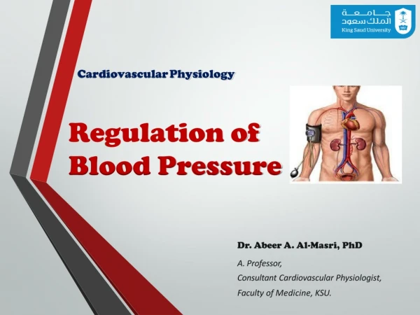
Cardiovascular Physiology Properties of the Cardiac Muscle
Properties of the cardiac muscle: • Excitability • Conductivity • Contractility • Rhythmicity
Properties of the cardiac Muscle I. Excitability (Irritability)
I. Excitability (Irritability): =the ability of cardiac ms to respond to adequate stimuli by generating an action potential followed by a mechanical contraction.
Relation between the action potential & the mechanical response ■The mechanical response consists of contraction (systole) & relaxation (diastole). ■Cardiac ms begins to contract few milliseconds after the AP begins, & continues to contract until few milliseconds after the AP ends. ■Duration of contraction: 0.2 sec in arial muscle, & 0.3 sec in ventricular muscle.
Relation between the action potential & the mechanical response(continued) ■ Diastole begins at the end of the plateau. ■ 2nd rapid repolarization is completed at about the middle of diastole.
Action potential of ventricular muscle ■Ventricular ms has a RMP of –90 mV. ( –85 to –95mV). ■The trans-membranous AP overshoots to a potential of ( +20mV).
AP of ventricular muscle(continued) Phase 0 = Rapid depolarization. Phase 1 = Rapid repolarization/ 1st rapid repolarization. Phase 2 = A plateau. Phase 3 = Slow repolarization/ 2nd rapid repolarization. Phase 4 = Complete repolarization. ■Trans-membranous AP of ventricular ms is characterized by presence of 5 phases.
AP of ventricular muscle(continued) 1 2 3 0 4 Phase 0= Rapid depolarization. ■opfast Na+ channels Na+ influx. Phase 1= Rapid repolarization/ 1st rapid repolarization. ■cls Na+ channels, K+ permeability, w Cl- influx. Phase 2= A plateau. ■op slow Ca2+ channels (slow Ca2+ Na+ channels) Ca2+ influx, w slow op K+ channels. Phase 3= Slow repolarization/ 2nd rapid repolarization. ■cls slow Ca2+ channels, w K+ permeability K+ efflux. Phase 4= Complete repolarization. ■actv Na+ K+ pump 2K+ in/ 3Na+ out.
Excitability changes during the action potential: ■ Passes through 3 periods: 1. Absolute refractory period (ARP) 2. Relative refractory period (RRP) 3. Dangerous period (supranormal period)
1. Absolute refractory period (ARP): ■The excitability of cardiac ms is completely lost during this period, i.e. doesn’t respond to 2nd stimulus. ■V. long. ■Occupies the whole period of systole. • ■Corresponds to the period of depolarization (phase 0), • & the first 2 phases of repolarization. • ■Ht can’t be tetanized (continuous contraction), as its • ARP occupies the whole contraction phase.
2. Relative Refractory Period (RRP): ■ The excitability of cardiac ms is partially recovered during this period, i.e. stronger stimuli than normal are required to excite the ms. ■Occupies the time of diastole. ■Corresponds to the 3rd phase of repolarization. • ■ Can be affected by the HR, temp., bacterial toxins, • vagal stimulation, sympathetic stimulation & drugs.
3. Dangerous Period (Supranormal): • ■ The excitability of cardiac ms is supranormal just • at the end of the AP, i.e. weaker stimuli than normal • can excite the ms. • ■? result in ventricular fibrillation.
Factors affecting myocardial excitability: 1. Cardiac innervation. 2. Effect of ions concentration in ECF. 3. Physical factors. 4. Blood flow. 5. Chemical factors (drugs).
Factors affecting myocardial excitability(continued) 1. Cardiac Innervation: ■ Sympathetic NS excitability. ■ Parasympathetic NS (vagus) excitability. 2. Effect of ions concentration in ECF: ■ Ca2+ excitability. ■ K+ excitability. 3. Physical factors: ■ temperature excitability. ■ temperature excitability.
Factors affecting myocardial excitability(continued) 4. Blood flow: ■Insufficient bl flow to cardiac ms excitability & myocardial metabolism for 3 reasons: (1) lack of O2, (2) excess accumulation of CO2, & (3) lack of sufficient food nutrients. 5. Chemical factors (drugs): ■Digitalis excitability.
Properties of the cardiac Muscle II. Conductivity
II. Conductivity: = the ability of cardiac ms fibers to conduct the cardiac impulses that are initiated in the SA-node (the pacemaker of the heart).
The direction of the impulse: ■The impulse is conducted: 1st Atrial spread ■ from SA-node conductive tissue ventricles. 2nd Ventricular spread ■ from apex of the heart base, via Purkinje fibers to the endocardial surface of ventricles.
The direction of the impulse(continued) N.B. LBB starts before RBB, as LV wall is thicker so the impulse needs more enough time to reach. Accordingly both ventricles will contract together.
Conduction of Impulse ■ APs from SA node spread quickly at rate of 0.8 - 1.0 m/sec. ■ Time delay occurs as impulses pass through AV node. • Slow conduction of 0.03 – 0.05 m/sec. ■ Impulse conduction as spread to Purkinje fibers at a velocity of 5.0 m/sec. • Ventricular contraction begins 0.1–0.2 sec. after contraction of the atria.
The conduction velocities of the impulse: SA-node 0.05 m/sec. AV-node 0.01 m/sec. … (slowest) Bundle of His 1.00 m/sec. Purkinje fibers 4.00 m/sec. …. (fastest) Atrial & Ventricular muscles 0.3 to 0.4 m/sec.
The conduction velocities(continued) ■The slowest conduction velocity in AV-node: ■ because it has few no. of intercalated discs. ■ Importance: to allow sufficient time for ventricles to be filled w bl before they contract. ■The fastest Conduction velocity in Purkinje fibers: ■ Importance: to allow the 2 ventricles to contract at the same time simultaneously.
Factors affecting myocardial conductivity: 1. Cardiac innervation. 2. Effect of ions concentration in ECF. 3. Physical factors. 4. Blood flow. 5. Chemical factors (drugs).
Factors affecting myocardial conductivity(continued) 1. Cardiac Innervation: ■ Sympathetic NS conductivity. ■ Parasympathetic NS (vagus) conductivity. 2. Effect of ions concentration in ECF: ■ Ca2+ conductivity. ■ K+ conductivity. 3. Physical factors: ■ temperature conductivity. ■ temperature conductivity.
Factors affecting myocardial conductivity(continued) 4. Blood flow: ■Insufficient bl flow to cardiac ms conductivity & myocardial metabolism for 3 reasons: (1) lack of O2, (2) excess accumulation of CO2, & (3) lack of sufficient food nutrients. 5. Chemical factors (drugs): ■Digitalis conductivity.
Properties of the cardiac Muscle III. Contractility
III. Contractility: = the ability of the cardiac muscle to convert chemical energy into mechanical work.
Contractility(continued) ♥Myocardial fibers have ‘Functional syncytium’ & NOT‘anatomical syncytium’, because they present in contact but NOT in continuity. ♥Strength of myocardial contraction determines the heart pumping power. ♥Mechanism of contraction depends on the contractile filaments, which contain the protein molecules (actin & myosin).
Excitation-Contraction Coupling in Heart Muscle = is the mechanism by which AP causes myofibrils of cardiac ms to contract. ♥ When AP passes over cardiac ms membrane, AP also spread to interior of cardiac ms fiber along membranes of transverse (T) tubules. ♥ Depolarization of myocardial cell stimulates opening of Ca2+ channels in sarcolema. • Ca2+ diffuses down gradient into cell through T tubules. • Stimulates opening of Ca2+-release channels in SR. • Ca2+ binds to troponin & stimulates contraction (same mechanisms as in skeletal ms).
Excitation-Contraction Coupling(continued) ♥ At the end of plateau of cardiac AP, i.e. during repolarization, ■ Ca2+ in sarcoplasm is rapidly & actively transported & pumped out of the cell via a Na+- Ca2+- exchanger, back into both SR & T tubules. ■ Resulting in cessation of the contraction until new AP occurs.
Factors affecting myocardial contractility: (Inotropic effectors) 1. Cardiac innervation. 2. Oxygen supply. 3. Calcium & potassium ions concentration in ECF. 4. Physical factors. 5. Hormonal & chemical factors (drugs). 6. Mechanical factors.
Factors affecting myocardial contractility(continued) 1. Cardiac Innervation: ■ Sympathetic NS force of contraction. ■ Parasympathetic NS (vagus) atrial force of contraction w no significant effect on ventricular ms.
Factors affecting myocardial contractility(continued) 2. Oxygen supply: ■ Hypoxia contractility. 3. Calcium & potassium ions concentration in ECF: ■ Ca2+ contractility. ■ K+ contractility. 4. Physical factors: ■ Warming contractility. ■ Cooling contractility.
Factors affecting myocardial contractility(continued) 5. Hormonal & chemical factors (drugs): ■ +ve inotropics: (Adrenaline, noradrenaline, alkalosis, digitalis, Ca2+, caffieen,…) ■ -ve inotropics: (Acetylcholine, acidosis, ether, chloroform, some bacterial toxins (e.g. diphtheria toxins), K+, …)
Factors affecting myocardial contractility(continued) 6. Mechanical factors: a. Cardiac ms. obeys ‘all or none law’: i.e. minimal or threshold stimuli lead to maximal cardiac contraction, because cardiac ms. behaves as a syncytium.
Factors affecting myocardial contractility(continued) b. Cardiac ms. can’t be stimulated while it is contracted, because its excitability during contraction is zero due to long ARP, so it can’t be tetanized. c. Cardiac ms. can perform both isometric & isotonic types of contractions.
Factors affecting myocardial contractility(continued) d. Starling’s law of the heart: ■ “Length-tension relationship” ‘Within limits, the greater the initial length of the fiber, the stronger will be the force of its contraction; However, overstretching the fiber as in heart failure its power of contractility decreases’ i.e. within limits, the power of contraction is directly proportional to the initial length of the ms. ■Cardiac ms accommodates itself (up to certain limit) to the changes in venous return.
Factors affecting myocardial contractility(continued) e.Cardiac ms shows staircase phenomenon (gradation), if providing all other conditions kept constant. i.e. if an isolated heart is stimulated by successive equal & effective stimuli, the 1st few contractions show a gradual in the magnitude of contraction.
Properties of the cardiac Muscle IV. Rhythmicity (Automaticity)
IV. Rhythmicity (automaticity): = the ability of cardiac ms to contract in a regular constant manner w/out nerve supply. ♥ It’s myogenic in origin (i.e. not neurogenic). ♥ Its initiated by the ‘pacemaker’ of the ht, the SA- node.
The pacemaker of the heart: =the SA- node. ♥Contains the P- cells, which are probably the actual pacemaker cells. ♥ Has the fastest rhythm (rate of discharge) of all parts of the heart, 90 impulses/min. its fibers have an unstable RMP. ♥ Has spontaneous (w/out stimulation) depolarization, up to firing level. ?
Pacemaker potential: ♥Its RMP is ( -60 mV). ♥ Pacemaker tissue is characterized by unstable membrane potential, Prepotential. ? -6
Pacemaker Prepotential: ? ♥Due togradual state of depolarization: ■ Steady in K+ permeability ( K+ efflux), leading to intracellular negativity. ■ Causing spontaneous leakage of membrane to Na+w/out stimulation. (-60 mV to -55 mV). ■ Which causes op of voltage gated transient Ca2+ channels, leading to some Ca2+ influx. (-40 mV). -6
Pacemaker Action potential (AP) -6 ♥Pacemaker Depolarization: • Opening of long lasting (fast) Ca2+ channels. • More Ca2+ influx till reaching the potential, i.e. firing level point leading to depolarization. • Opening of VG Na+ channels ? also contribute to the upshoot phase of the AP.
Pacemaker Action potential (AP)(continued) -6 ♥Pacemaker Repolarization: • Opening of VG K+ channels. • K+ diffuses outward (efflux), … (so +vity will go out of cell). ♥Pacemaker Hyperpolarization: ■ excessive K+ effllux, (This will lead to hardship of K+ efflux in 2nd depolarization). • Ectopic pacemaker: • Pacemaker other than SA node: • If APs from SA node are prevented from reaching these areas, these cells will generate pacemaker potentials.