Download

1 / 60
620 likes | 925 Views
Learn about the general organization and function of the digestive system for obtaining metabolites. Explore cellular structures and functions in the digestive system efficiently.

E N D
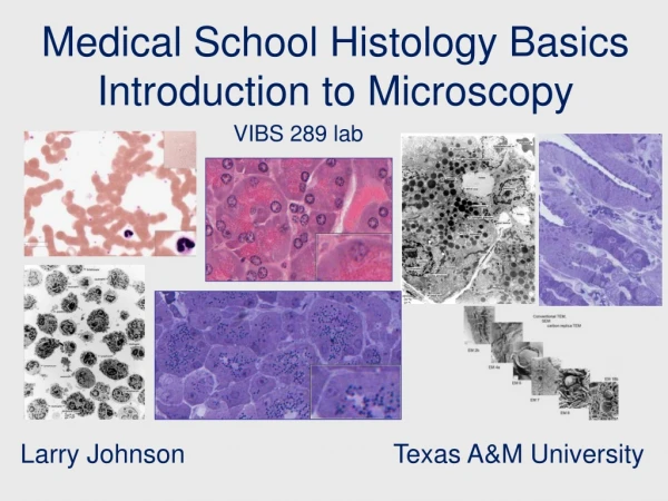
Medical School Histology Basics Digestive System VIBS 289 lab Larry Johnson Texas A&M University
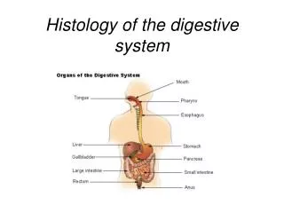
Objectives To understand the general organization of organs of the digestive system and how they function to obtain metabolites necessary for growth and energy for the body, yet maintain a barrier between the environment and the internal milieu of the body To identify and describe functions of cellular structures, cells, and groups of cells in the digestive system.
Function of the Digestive System Movement of food Secretion of digestive juices Absorption of digested foods, water, and electrolytes
Adaptation of G.I. Tract for Specific Function FunctionOrgan Simple passage from one part Esophagus to another Storage of food or feces Stomach or distal colon Digestion Stomach, small intestine Absorption of end products Small intestine, proximal colon
32409 Small intestine Large intestine Small intestine
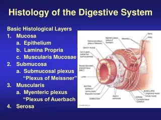
32409 General Structure of the Digestive Tract Epithelium Lamina propria Muscularis mucosa Submucosa Muscularis externa Serosa
Small intestine 146 General Structure of the Digestive Tract Epithelium Lamina propria Submucosa Muscularis externa Serosa 32409 Small intestine
Large intestine 153 General Structure of the Digestive Tract Epithelium Lamina propria Muscularis mucosa Submucosa Muscularis externa Large intestine Stomach 145
Large intestine Mesothelium Muscularis externa 153 145 153 Stomach Muscularis externa Inner, thicker circular layer Outer longitudinal layer of the muscularis externa Serosa
Histo 51 Filiform Papillae Non-keratinized stratified squamous epithelium. Skeletal muscle, Mucus and Serous glands,
Slide #12 (1101). Tongue, rabbit. Filiform papillae Skeletal muscle Foliate papillae that possess Taste buds Serous glands
Esophagus Non-keratinized stratified squamous epithelium. Histo 29 Muscularis externa Sub-mucosal glands Skeletal muscle
137 Esophagus – skeletal and smooth muscle epithelium Lamina propria Muscularis mucosa Submucosa Muscularis externa Serosa
Esophagus Sub-mucosal 242 Mucus and Serous glands Submucosa Lamina propria Muscularis mucosa Non-keratinized stratified squamous epithelium Skeletal muscle, Muscularis externa If outer layer is not covered by mesothelium = adventitia Submucosa Perichondrium
137 Esophagus Muscularis externa of the upperesophagus is composed mostly of skeletal muscle The muscularis externa in middle to lower esophagus is composed mostly of smooth muscle.
437 Cardio-esophageal junction Sratified squamous Luminal epithelium changes from stratified squamous to simple columnar epithelium in the cardiac region
The stomach have no goblet cells, no brush border on surface cells, and no villi.
Dog cross section of body Stomach Liver Pancreas Esophagus Intestine
145 Fundic stomach Mucosa Antibody-producing plasma cells Gastric pits Submucosa Ganglion cells of the Auerbach's plexus regulate the muscularis externa Submucosa Ganglion cells of the Meissner's plexus regulates muscularis mucosa Muscularis externa
145 Fundic stomach Surface mucous cells Mucous neck cells Gastric pits and gastric glands Chief cells and parietal cells Enteroendocrine cells
Mucus neck cells Fundic stomach, rabbit (toluidine blue) 244 Enteroendocrine cells Chief cells Parietal cell
EM 15 Parietal cell produces • HCl • Bicarbonate • Intrinsic factor for vitamin B12 absorption by gut: needed in red blood cell formation
243 Fundic stomach, monkey (PAS) Mucous neck cells Parietal cells Chief cells Enteroendocrine cells Surface mucous cells
244 Fundic stomach, rabbit (toluidine blue) Parietal cells Gastric pits Surface mucous cells Mucous neck cells Lumen Enteroendocrine cells Lumen
PAS H&E Surface mucus cells EM 16 • Granules of surface mucous cells • Mitochondria • Nuclei • Lumen Toluidine blue
Large granules of chief cell Granules of an argentaffin cell EM 14 • Large granules of chief cell • Granules of an argentaffin cell • Lamina propria • Nuclei
141 Pyloric stomach, monkey (PAS) Pyloric glands Pyloric glands of the stomach contain mucous cells Surface mucus cells
147 Pyloroduodenal junction, baboon Intestine Lymphoid nodule Stomach Muscularis mucosa Muscularis externa Lamina propria
147 Pyloroduodenal junction, baboon Submucosa Gastric glands Stomach Muscularis mucosa Muscularis externa Gastric pits Intestine Villi Lamina propria Crypts of Lieberkühn (intestinal glands). Stomach intestine Junction Goblet cells and intestinal absorptive cells with a brush border
146 Duodenum, monkey Goblet and absorptive cells, Enteroendocrine cell Crypts of Lieberkühn Muscularis mucosa Lamina propria. Submucosa Submucosal Brunner's glands.
152 Duodenum Absorptive cells Brunner’s glands Goblet cells Paneth cell Enteroendocrine cell Intestinal villus
Mucus of goblet cells and the carbohydrates in the brush border are PAS positive for sugars 447 Small intestine Intestinal villus Intestinal absorptive cell Brush border Macrophages Intestinal absorptive cell Brush border Goblet cells Central lacteal 249 PAS
EM 17 Basal portion of intestinal absorptive cell • Plasma cell • Lymphocyte • Smooth muscle • Intestinal absorptive cell • Macrophage • Lumen of capillary • Pericyte of capillary
Goblet cell EM 4. Apical portion of intestinal absorptive cell • Microvilli of brush border • Droplets of goblet cell • Terminal web • Lipid in SER • lumen
Basal portion of intestinal absorptive cell • Mitochondria • Nuclei of intestinal absorptive cell • Smooth muscle of muscularis mucosa • Basal lamina
EM 4b Brush border of intestinal absorptive cells Mitochondria
EM 4c Intestinal absorptive cells in cytoplasm just above the nucleus Nucleus
Epithelium Smooth muscle cells Meissner’s plexus cell bodies 32409 (Auerbach's plexus) Nerve cell bodies
152 Duodenum (small intestine) Auerbach's plexus, found in between the circular and longitudinal smooth muscle layers in small intestine Auerbach's plexus Nerve cell bodies
148 Meissner’s plexus cell bodies in submucosa Ileum Paneth cell Submucosa Small intestinal villi
250 Argentaffin cells of Ileum, monkey Enteroendocrine cells also called Argentaffin cells Paneth cell
Compare luminal surfaces of the small and large intestines NO Villi small intestines Villi large intestines