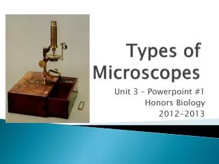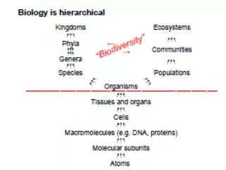
Microscopes—tools for studying biodiversity
Microscopes are essential for studying biodiversity, enabling scientists to detect and distinguish between various organisms by analyzing the intricate structure of cells, tissues, and organs. Simple microscopes utilize a single lens to magnify objects, while compound microscopes feature multiple lenses for higher magnification. Advanced electron microscopes provide enhanced resolution, allowing for the visualization of fine structures and even viruses. This exploration of microscopy also highlights historical contributions from scientists like Robert Hooke and Antonie van Leeuwenhoek, who paved the way for microbiology.


Microscopes—tools for studying biodiversity
E N D
Presentation Transcript
Microscopes—tools for studying biodiversity • detect, distinguish between organisms • study structure of cells, tissues, organs • determine locations of molecules
simple microscope • 1 lens (eg magnifying glass) • bends light so object appears larger
compound microscope • >1 lens • mag multiplies • so do optical problems
optical terms • magnification: how large appears compared to actual size • resolution (resolving power): closest distance 2 objects can be, & still be distinguished as separate obj. • resol dep on wavelength () • shorter better. electron microscopes
lens resolution comparison • human eye 0.2mm ruler in lab • light scope 0.2m 1000x better than eye • electron scope ~ 2 nm 100x better than LM 100,000x better than eye • = micro • LM = light scope • EM = electron scope
scale • 1mm = 1000 m = 1 million nm • human hair ~1/10 of mm = 100 m • electron microscopy reveals viruses! 20 – 90 nm • 1990s: 50 million/mL of seawater, soil • Fig 6.2, p 95
image terms • photograph--no scope • photomicrograph--photo taken though scope • light micrograph • electron micrograph
LM • Advantage: living cells & organisms • colors (pigments) • movement • focus through depth of specimen
EM • disadvantage: specimens dead • electrons from filament • in vacuum • focus with electromagnetic lenses
TEMtransmission electron microscope • electrons go through specimen • make shadow • fine structure inside of cells • many sections for 3-D structure
TEM • can locate molecule w/antibody attached to gold particle
SEM scanning electron microscope • electron beam scans specimen • specimen electrons collected • surface of object • 3-D view
LM contrast • pigments • stains • brightfield--standard bright background • special techniques (Fig 6.3, p 96) • polarized light • darkfield • differential interference contrast (dic) • phase contrast
fluorescence microscopy • UV light source • specimen emits light • autofluorescence • tagged antibodies • reporter proteins
Confocal microscopy • laser light source • optical sections • no out of focus blur
discovery of cells • 1665 English scientist Robert Hooke • first saw (cork) cells [cell = room] • compound microscope • (30x mag) • Royal Society, London Hooke’s drawing of cork cells in Micrographia http://www.gutenberg.org/files/15491/15491-h/15491-h.htm section 18, plate 11
Hooke’scompound microscope http://www.arsmachina.com/hooke.htm
scientific literature • primary lit. — original report of research • intro, materials & methods, results, discussion • peer-reviewed • secondary lit. — report about primary lit. • ProcRSocL, Nature, PNAS, Science
discovery of microorganisms • 1674 Dutch merchant Antony van Leeuwenhook • first saw “animalcules” • protists, and later bacteria • simple microscope w/great lens • 275-295x mag http://www.ucmp.berkeley.edu/history/leeuwenhoek.html
discovery of microorganisms • 1676 Leeuwenhook wrote to Royal Society @ singled celled organisms • Hooke confirmed • first published bacteria • voucher specimens http://www.ucmp.berkeley.edu/history/leeuwenhoek.html
Leeuwenhoek’s microscope light micrograph of red blood cells photographed through this scope http://www.brianjford.com/wav-mict.htm