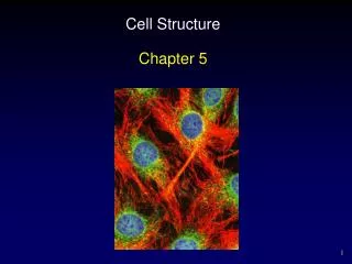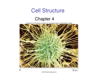
Cell Structure
Light Microscopy. Types of microscopes used to view microbes: - Light microscopes: (1) Bright field (2) Phase contrast (3) Dark-field (4) Fluorescence - Electron microscopes: (1) Scanning (SEM) (2) Transmission (TEM).


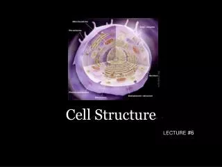
Cell Structure
E N D
Presentation Transcript
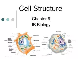
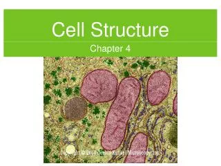
1. Chapter 4 Cell Structure/Function
2. Light Microscopy Types of microscopes used to view microbes: - Light microscopes: (1) Bright field (2) Phase contrast (3) Dark-field (4) Fluorescence - Electron microscopes: (1) Scanning (SEM) (2) Transmission (TEM)
3. Bright-Field Microscopy Consists of 2 series of lenses (objective lens and ocular lens) that resolve the image.
Specimens are made visible because of the differences in contrast between them and the surrounding medium. Why then are many bacterial cells difficult to see with a bright-field microscope?
4. Magnification vs. Resolution Components: - Magnification: can be increased without limit. - Resolution: the ability to distinguish two points as separate, cannot be increased without limit � why?
Resolution of light microscopy vs. electron microscopy: - Light microscopy: 0.2�m (200 nm) What does this number mean? - Electron microscopy: 1000X greater � so, what does this make it?
Total magnification = magnification of objective lens x magnification of the ocular lens Resolution cannot be increased without limit because it is dictated by the physical properties of light.
Electron microscopy resolution = 1000X 0.2 �m (200 nm) = 0.0002 � (0.2 nm).
The measure of resolution means that two objects closer together than this value cannot be distinguished as being separate.Resolution cannot be increased without limit because it is dictated by the physical properties of light.
Electron microscopy resolution = 1000X 0.2 �m (200 nm) = 0.0002 � (0.2 nm).
The measure of resolution means that two objects closer together than this value cannot be distinguished as being separate.
5. Magnification vs. Resolution (continued) Max magnification of compound light microscopy = 1500 X
Resolution = function of wavelength of light used and the numerical aperture of the objective lens (measure of light-gathering ability).
Most oculars = 10-15 X, most objectives = 10-100X
The 100X objective is typically called the oil immersion objective because high-grade optical oil is used with this lens to increase the light-gathering ability of the lens. How does this work? Oil immersion works by allowing rays emerging from the specimen at higher angles to be collected and viewed, whereas these rays would otherwise be lost to the objective lens.Oil immersion works by allowing rays emerging from the specimen at higher angles to be collected and viewed, whereas these rays would otherwise be lost to the objective lens.
6. Staining Purpose: Increase contrast for bright-field microscopy.
Positively charged (cationic) dyes: bind to negatively charged cellular components (nucleic acids, acidic polysaccharides, cell surfaces), ex. methylene blue, crystal violet, safranin.
General procedure: slide containing a dried suspension of microbes is flooded with a dilute solution of the dye for a minute or two, rinsed in water, and blotted dry. These are often observed with the oil immersion lens.
7. Differential Staining Does not stain all kinds of cells equally.
Most important differential stain = Gram stain. It is the often the first step in identifying unknown bacteria. Do you remember what the stain binds to in the cell? What color do gram-positive cells stain? What about gram-negative cells?
Refer to Fig. 4.4 in the text. Gram stain stains different structures of the cell wall. Gram-positive cells stain purple and gram-negative cells stain pink. Gram stain stains different structures of the cell wall. Gram-positive cells stain purple and gram-negative cells stain pink.
8. Phase Contrast Microscopy A special ring in the objective lens causes a dark image to be formed on a light background.
Advantages: developed to increase contrast between cells and the surrounding medium without staining � why not just stain them? Drying and fixing as well as staining can cause changes in the cells and introduce artifacts. Artifacts = foreign structures introduced that aren�t there naturally.Drying and fixing as well as staining can cause changes in the cells and introduce artifacts. Artifacts = foreign structures introduced that aren�t there naturally.
9. Dark-Field Microscopy Lighting has been modified to reach the specimen from the sides only.
The only light reaching the lens is light scattered by the specimen
Specimen appears light on a dark background.
Advantages: has higher resolution than bright-field or phase contrast microscopes, good for viewing flagella and motility.
10. Fluorescence Microscopy Used to visualize specimens that fluoresce � what does this mean?
Cells fluoresce because: - they contain naturally fluorescent substances, ex. chlorophyll or other fluorescing components (what is this called?) - they have been treated with a fluorescent dye.
Uses: clinical diagnostic microbiology and microbial ecology. Fluoresce: emit light of one color when light of another color shines upon them.
Autofluorescence.Fluoresce: emit light of one color when light of another color shines upon them.
Autofluorescence.
11. 3-D Imaging: Differential Interference Contrast Microscopy A form of light microscopy that employs a polarizer to polarize light, which passes through a prism to separate the light, followed by an objective lens that recombines the light. The combined beams are not in phase, but create an interference effect, which intensifies differences in cell structure.
Structures in cells such as a nucleus, spores, vacuoles, granules take on a 3-D appearance.
Advantages: useful for observing unstained cells.
12. 3-D Imaging: Atomic Force Microscopy Tiny stylus is positioned close to a specimen.
Weak repulsive atomic forces are established between the probe and the specimen.
The pattern is monitored by detectors and images are created that look like SEM images.
Advantages: specimen prep. for AFM is like that for light microscopy instead of SEM. Also, hydrated specimens can be viewed, which cannot be done by SEM.
13. 3D-Imaging: Confocal Scanning Laser Microscopy Computerized microscope that couples a laser light source to a light microscope.
Relatively think specimens can be observed in terms of not only the surface, but the layers as well, by adjusting the plane of focus of the laser beam.
Cells may be stained with fluorescent dyes or false color images can be generating by adjusting the microscope so that different layers take on different colors.
Advantages: images stored digitally, used in microbial ecology, and for thick specimens.
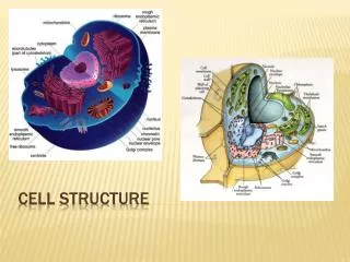
14. Transmission Electron Microscopy (TEM) Used to study internal structures of a cell.
Electrons are used instead of light rays, electromagnets are used instead of lenses, system operates under high vacuum.
Requires special sample prep.: fixing, precise thin sectioning (20-60 nm slices), and staining with high atomic weight stain, ex. osmic acid, uranium salts, lead � they scatter electrons well, improving contrast.
Advantages: can see things as small as proteins and nucleic acids.
15. Scanning Electron Microscopy (SEM) Used to view external features of a specimen.
Special sample prep.: specimen has to be coated with a thin film of a heavy metal, ex. gold.
Electron beam is directed onto a specimen and scans back and forth across it. Electrons scattered by the metal are collected, producing an image.
Advantages: can be used to view intact cells, large specimens, magnifies from 15 X to 100,000 X.
Both TEM and SEM: can take a picture of the image called an electron micrograph.
16. Cell Morphology What does morphology mean?
3 of the most common shapes:
- sphere-shaped = coccus (pl. = cocci)
- rod-shaped = bacillus (pl. = bacilli)
- spiral-shaped = spirillum (pl. = spirilli)
Arrangements: Cells may be arranged in chains, clusters, etc.
Unusual shapes: spirochetes, appendaged, filamentous. What makes each unusual? Morphology = shape.
Spirochetes = tightly spiraled, appendaged = cells extended into long tubes or stalks, filamentous = form long thin cells or chains of cells.Morphology = shape.
Spirochetes = tightly spiraled, appendaged = cells extended into long tubes or stalks, filamentous = form long thin cells or chains of cells.
17. The Significance of Being Small Compare sizes:
- Prok.: 0.1-0.2�m to 50�m in diameter
- E. coli: 1 X 3 �m
- Euk.: 2-200�m
The small size of prok. affects their biological properties: rate at which nutrients and waste products pass into and out of a cell ? affects cellular metabolic and growth rates and is inversely proportional to size.
Transport rates are to some degree a function of membrane surface area available, relative to cell volume.
18. The Significance of Being Small (continued) Ex. sphere: V = 4/3?r, SA = 4?r, surface:volume = 3/r
? a cell with a smaller r has a higher S/V ratio than a larger cell and thus can have more efficient exchange of nutrients with its surroundings. This results in faster growth rates and larger cell populations for smaller cells. What can this result in?
How do the membrane-bound organelles of euk. impact this concept?
Nanobacteria? Major physiological changes in an ecosystem in a short amount of time.
The existence of nanobacteria, 0.1�m in size, has been proposed . Most say this is too small to contain the essential molecules of life.Major physiological changes in an ecosystem in a short amount of time.
The existence of nanobacteria, 0.1�m in size, has been proposed . Most say this is too small to contain the essential molecules of life.
19. Cytoplasmic (Cell) Membrane Phospholipid bilayer � refer to Fig. 4.15 and 4.16 of the text.
Integral membrane proteins (refer to Fig. 4.17 of the text) vs. peripheral membrane proteins � what�s the difference? What are the properties of each?
What kinds of things happen at the outer surface of the cell membrane?
What kinds of things happen at the inner surface of the cell membrane?
What is the fluid mosaic model of the cell membrane? Integral membrane proteins: have middle hydrophobic portion spanning the membrane, flanked by a hydrophilic portion extending out of the membrane to the outside of the cell and another hydrophilic portion extending out of the membrane on the cytoplasmic side.
Peripheral membrane proteins: lipoproteins = proteins that have a lipid tail, which attaches them to the membrane.
Outer surface: makes contact with variety of proteins that bind substrates or process large molecules for transport into the cell.
Inner surface: Energy-yielding reactions.
Fluid mosaic model: show Bio 113 video clip.Integral membrane proteins: have middle hydrophobic portion spanning the membrane, flanked by a hydrophilic portion extending out of the membrane to the outside of the cell and another hydrophilic portion extending out of the membrane on the cytoplasmic side.
Peripheral membrane proteins: lipoproteins = proteins that have a lipid tail, which attaches them to the membrane.
Outer surface: makes contact with variety of proteins that bind substrates or process large molecules for transport into the cell.
Inner surface: Energy-yielding reactions.
Fluid mosaic model: show Bio 113 video clip.
20. Cytoplasmic (Cell) Membrane (continued) Euk.: sterols in cell membrane, sterols = ridid molecules that strengthen and stabilize the phospholipid bilayer.
Bacteria: contain hopanoids instead of sterols, except the methanotropic bacteria and mycoplasmas.
Archaea: contain ether bonds instead of ester bonds between glycerol and side chains, contain isoprene side chains instead of fatty acids. Some Archaea contain a monolayer instead of a bilayer. Which have this and what advantage is it? Refer to Fig. 4-19 and 4-20 in the text. Hyperthermophilic Archaea contain a monolayer because it is not as easily pulled apart.Hyperthermophilic Archaea contain a monolayer because it is not as easily pulled apart.
21. Cell Membrane: Semi-Permeable Membrane Prevents leakage of cytoplasmic constituents out of the cell.
Prevents foreign things from getting in (for the most part).
Site of many proteins that transport things into and out of the cell.
Can be energetically charged, keeping H+ ions on one side and OH- ions on the other side. This charge separation = a form of energy, like the potential energy of a battery. The membrane in this state is called a proton motive force and can be used to drive energy-requiring rxns. such as transport.
22. Cell Membrane: Semi-Permeable Membrane (cont.) Inside the cell: aqueous soln. of salts, sugars, amino acids, vitamins, coenzymes, etc.
Cell membrane = hydrophobic barrier.
Some small hydrophobic molecules pass through the membrane by diffusion, but hydrophilic and charged molecules must be specifically transported, ex. H+ and OH-.
Water is small enough to pass through the membrane, but is often transported by aquaporins.
23. Transport Proteins Ferry things across the membrane in a very specific fashion, and often do so against the concentration gradient b/c the conc. of nutrients in nature is often very low.
Show a saturation effect at which point the rate of uptake is maximum and will not increase beyond that rate.
3 types of transport systems - all require energy (proton motive force, ATP, etc.): 1. Only membrane-spanning component 2. Membrane-spanning component + periplasmic-binding component. 3. Series of proteins that cooperate, ex. phosphotransferase system.
24. Transport Systems - Refer to Fig. 4-23 and 4-24 of the text
25. Types of Simple Transporters: 1. Uniporters: transport a molecule in one direction. 2. Symporters: transport a molecule along with another substance, ex. H+. 3. Antiporters: transport a molecule in one direction while transporting another molecule in the opposite direction.
26. Lac Permease: A Simple Transporter Lactose is transported by Lac permease - what is this?
What energy enables this to occur? What happens when that energy is depleted?
What is lactose exactly and of what use is lactose to the cell?
What type of simple transporter is Lac permease?
27. Group Translocation Process in which substance being transported is chemically altered as it is being transported.
Ex. sugars (glucose, fructose, mannose) that are phosphorylated by the phosphotransferase system.
Phosphate group is supplied by phosphoenolpyruvate (PEP).
28. Group Translocation (cont.) Phosphorylation of glucose = 1st step in pathway for its metabolism.
Phosphate is passed along the series of proteins 1st and then attached to glucose.
29. ABC System Dep. on periplasmic binding proteins.
Periplasmic space = space that gram-neg. bacteria have between cytoplasmic membrane and lipid-rich outer membrane called the periplasm.
Components: 1. Mem.-spanning protein 2. Periplasmic-binding protein 3. ATP-hydrolyzing protein.
30. ABC System (cont.) ABC = ATP-binding cassette
200 diff. ABC systems identified = family of related proteins.
Periplasmic-binding proteins have very high affinity for their substrates.
Gram-positive bacteria have similar mechanism, but the the specific binding proteins are not mobile (anchored to membrane).
31. Protein Export Tranlocases transport large molecules, ex. proteins, out of the cell = secretory system.
Proteins contain special signals that designate them for export.
Uses: - hydrolytic enzymes, ex. amylase, cellulase, excreted into environment to begin extracellular digestion. - toxins excreted by pathogenic bacteria - why to pathogenic bacteria do this (what is the advantage)?
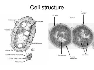
32. Cell Wall: PG, etc. Typical bacteria contain a turgor pressure similar to an automobile tire, and therefore need to be able to withstand such pressure ? cell wall (also gives shape and rigidity to the cell.
2 major groups: 1. gram-positive - thick single layer = peptidoglycan (PG) 2. gram-negative - multilayered, rigid layer = PG, lipopolysaccharide (LPS) layer over that.
PG consists of: alternating units of ?-1,4 linked N-acetylglucosamine and N-acetylmuramic acid, and a small group of amino acids forming the glycan tetrapeptide, which forms peptide cross-links.
33. Cell Wall: PG, etc. (cont.) Only when PG is cross-linked does the cell wall have full strength.
Gram-postive cells tend to have an interbridge, while gram-negative cells do not.
Gram-positive bacteria: up to 90% of cell wall is PG + some teichoic acids.
Gram-negative bacteria: only about 10% of cell wall is PG, rest is usually the outer membrane.
34. PG (cont.) DAP: present in gram-negative bacteria, but not common in gram-positive bacteria.
NAM and DAP: never found in the cell walls of Archaea or Eukarya; only Bacteria have PG.
Gram-positive Bacteria have acidic polysaccharides called teichoic acids or lipoteichoic acids embedded in their cell wall. TA�s impart a negative charge on the cell surface.
35. Gram-Positive Cell Wall
36. Lysozyme and Protoplasts PG can be destroyed, ex. by lysozyme: enzyme that breaks the ?-1,4 glycosidic bonds between NAG and NAM.
When these bonds are broken, water rushes in and causes the cell to swell and burst = lysis.
Lysozyme is found in animal secretions, ex. tears, saliva, etc. as a major line of defense against Bacteria.
Sucrose can be added to the medium to osmotically stabilize cells whose walls have been digested by lysozyme. These cells are called protoplasts.
What prok. can survive without cell walls, i.e., are natural protoplasts? Why are they able to do so?
37. PseudoPG - Archaea Contain NAG and N-acetyltalosaminuronic acid (instead of NAM).
Contain ?-1,3 linkages, instead of ?-1,4.
Species of Archaea exhibit a wide variety of cell wall types (pseudoPG and otherwise), but with rare exception, all contain a cell wall of some sort, which functions to prevent osmotic lysis and to define cell shape, as Bacterial cell walls do.
Archaea are naturally resistant to lysozyme, which destroys PG, and penicillin, which prevents the synthesis of PG.
38. Outer Membrane of Gram-Negative Bacteria Gram-negative cells contain a layer of PG, with a layer of lipopolysaccharide (LPS) or outer membrane over it.
LPS: consists of the core polysaccharide + the O-polysaccharide + lipid A.
The PG in the gram-negative CW is too thin to prevent passage of the alcohol into the PG (as opposed to the thick-PG-walled gram-positive CW), so the crystal violet is able to be washed out.
39. Endotoxin The outer membrane is often toxic to animals, ex. Salmonella, Shigella, Escherichia, etc.
Toxic properties are due to the lipid A portion of the LPS = endotoxin.
Nonpathogenic bacteria can have endotoxin activity.
40. Porins and the Periplasm Porins = channels for the entrance and exit of hydrophilic low-MW substances .
Porins can be specific or non-specific.
Porins are transmembrane proteins.
The outer membrane functions to keep certain enzymes outside the cytoplasmic membrane, but within outer membrane = the periplasm.
The enzymes in the periplasm = hydrolytic enzymes: initial degradation of food molecules, serve as binding proteins (transport substrates into the cell), and chemoreceptors (involved in the chemotaxis response).
41. Modes of Motility Most prok. are motile, though not all.
Flagella (sing. = flagellum): most common method
Gliding
Gas Vesicles
Purpose: new resources, opportunities, interactions.
Cost: energy expenditure.
42. Flagella Long, thin, attached to the cell at one end, can only be seen by special staining with a light microscope.
Positioning: - Polar: attached to one or both ends of a cell. - Lophotrichous: arranged in tufts at one end of a cell. - Peritrichous: inserted at many places around the cell surface.
Structure: - helically-shaped, have a characteristic wavelength, composed of protein subunits = flagellin. - composed of the filament + base (= hook + motor).
43. Flagella (cont.) Synthesis: - 40+ genes required for components and export/assembly of components. - individual flagellum grows from the tip (or cap), not the base.
Movement: Proton motive force drives the motor to rotate the flagellum ? cell moves 60 cell lengths/sec. vs. cheetah = 25 body lengths/sec.
44. Gliding Motility Slower than flagellar movement.
Cells are filamentous or rod-shaped.
Requires cells to be in contact with solid surface.
Ex. Myxococcus sp.
Mechanisms: - slime secretion (cyanobacteria): slime adheres to surface and pushes cells along - gliding proteins: provide ratcheting motion.
Refer to Fig. 4.44 (c) in the text for colonial morphology.
45. Behavioral Response: Chemotaxis What�s a gradient?
Motility mechanisms in cells guide the cells toward or away from gradients in the environment. These directed movements = taxes (sing. = taxis, ? not the kind you pay to the IRS).
How do cells sense gradients?
What is chemotaxis a response to?
Bacteria respond to temporal (as opposed to spatial) gradients of signal molecules as they swim along - what does this mean and how does this work?
46. Behavioral Response: Chemotaxis (cont.) How is bacterial movement in response to a signal characterized?
Bacteria move up a gradient (increasing in concentration) in response to an attractant and down a gradient (decreasing in concentration) in response to a repellent.
Changes in movement can be complex in microbes with peritrichous flagella or very simple in microbes with one flagellum. How does a microbe with one flagellum reorient itself? (What is Brownian movement?)
Chemotaxis is controlled at the genetic and biochemical levels, and involves chemoreceptors. How can chemotaxis be observed?
47. Behavioral Reponse: Phototaxis What is phototaxis?
Why do microbes phototax?
Phototaxis vs. scotophobotaxis?
Entire colonies can show phototaxis.
Mechanisms are thought to be similar to chemotaxis: photoreceptor is involved and similar signals control movement in response to sensing of light gradient.
Other taxes include aerotaxis = movement toward or away from oxygen, osmotaxis = movement toward or away from conditions of high ionic strength.
How does taxis relate to what all living cells have in common?
48. Fimbriae and Pili Structurally similar to flagella, but not involved with motility.
Fimbriae: shorter and more numerous than flagella, made of protein, function unknown for certain, but probably allow microbes to stick to surfaces, ex. pathogens to animal cells or form pellicles or biofilms.
Pili: similar structurally to pili, but longer and fewer in #, serve as receptors for viruses, involved in conjugation in prok. As well as attachement of pathogens to human tissues.
49. S-Layers Paracrystalline surface layers � have crystalline appearance (geometrical)
2-D array of protein
Basically all Bacteria and many Archaea have these.
= cell wall for some Archaea
Major function is unknown: acts as permeability barrier, may protect pathogenic bacteria against host defenses
50. Capsules and Slime Layers: Glycocalyx Many prok. secrete slimy or gummy material on surface.
Capsule or slime layer = glycocalyx = these types of polysaccharide layers.
Capsule = rigid, excludes particles.
Slime layer = not so rigid, does not exclude particles.
Glycocalyx functions: - attachment of pathogens to hosts - capsulated bacteria are equipped with a means for escape of destruction by phagocytic immune cells - resistance to desiccation (what�s that?)
51. Carbon Storage Polymers Functions of Granules: energy storage, reservoir of structural building blocks
Poly-?-hydroxybutyric acid (PHA): lipid-like, contained in inclusion bodies in prok., ex. granules, serve as C and energy storage.
Gycogen: glucose polymer, C and energy storage molecule produced by prok. and animal cells.
52. Other Granules and Inclusions Polyphosphate granules: store inorganic phosphate (Pi) for making nucleic acids and phospholipids.
Elemental sulfur granules: are the product of oxidation of reduced sulfur compounds for energy ? eventually reduced to sulfate and used up.
Magnetosomes: iron mineral magnetite, impart permanent magnetic dipole to a cell, cell responds to magnetic field, guide aquatic cells toward sediments, which are lower in oxygen (they like this).
53. Gas Vesicles Present in a # of prok. living in lakes and seas.
Give the cells buoyancy, motility.
Spindle-shaped.
Present in the cytoplasm.
Permeable only to gases.
Cells may contain a few to hundreds.
Made up of only proteins and only 2 types: GvpA (97%) � provides resistance to pressure - and GvpC (3%) � provides strength. Both are hydrophobic.
54. Endospores Process of forming = sporulation.
Resistant to heat, drying, radiation, harsh chemicals.
Commonly found in the soil.
Ex. include: Bacillus, Clostridium.
Structure: Exosporium, spore coats, cortex, core or spore protoplast. What chemical is specific to endospores?
Properties: contains only 10-30% of the water that the vegetative cell contains, enzymes are inactivated, pH is lowered. What 2 functions do small acid-sol. spore proteins _SASPs) have?
55. Endospore Formation and Back Again Sporulation involves a complex series of events of cellular differentiation.
When does sporulation occur?
Sporulation is genetically-directed.
It can take 8 hours and 200 genes.
What are the steps of conversion from an endospore back into a vegetative cell? What triggers this and how long does it take to occur?
What types of cells undergo sporulation? When the bacterium has run out of nutrients (nutrients have become limiting).
(1) Activation, (2) Germination, (3) Outgrowth. Activation is triggered by the return of nutrients and acceptable growing conditions and can occur quickly (in minutes). Germination also can occur within minutes and includes shedding of the endospore layers and protective properties. Outgrowth involves swelling due to water uptake, synthesis of new RNA, proteins, and DNA.
A sublineage of gram-positive bacteria undergo sporulation and include anaerobes, aerobes, phototrophs, and chemolithotrophs.
When the bacterium has run out of nutrients (nutrients have become limiting).
(1) Activation, (2) Germination, (3) Outgrowth. Activation is triggered by the return of nutrients and acceptable growing conditions and can occur quickly (in minutes). Germination also can occur within minutes and includes shedding of the endospore layers and protective properties. Outgrowth involves swelling due to water uptake, synthesis of new RNA, proteins, and DNA.
A sublineage of gram-positive bacteria undergo sporulation and include anaerobes, aerobes, phototrophs, and chemolithotrophs.