Download
1 / 49
570 likes | 762 Views
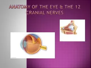
Learn about the 12 pairs of cranial nerves and their nuclei attachment in the brain stem and forebrain, functions, and pathways. Understand the afferent and efferent nerve nuclei in the brain stem.

E N D
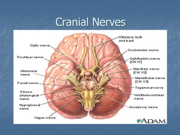
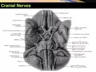
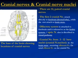
Cranial nerves & Cranial nerve nuclei : • There are 12, pairedcranial nerves. • The first 2 cranial Ns. attach directly to forebrain (frontal lobe) , while the rest attach to brain stem. • Olfactory system is attached to forebrain and is referred to as the limbic system, / optic N. also is discribed in visual pathway. • Cranial Ns. from 3 - 12 havenuclei (cranial N.nucluei) in the brain stem , receiving afferents Fs. Or send efferent Fs. as the cranial Ns. The base of the brain showing locations of cranial nerves
Superficial attachements of Cranial nerves : • Occulomotor & trochlear Ns. are attached to midbrain. • Trigeminal N. is attached to antero-lateral surface of pons. • Abducent, Facial & vestibulo-cochlear Ns. are lying between pons & M.O. from medial to lateral. • Hypoglossal N. is attached to antero-lateral sulcus of M.O. / but glossopharyngeal, vagus & accessory Ns. are attached to postero-lateral sulcus of M.O. The base of the brain showing locations of cranial nerves
Afferent Nerve Nuclei : • Fibres carrying general sensationfrom the headvia trigeminal N. terminate in a large trigeminal sensorynucleusthat extends the whole length of brain stem andcervical spinal cord. • Fibres carrying hearing & motion/positional sensevia vestibulo-cochlear N. terminate in cochlear & vestibularnuclei that are located in upper M.O. • Visceral afferents carrying tastesensationvia facial & glossopharyngeal Ns., terminate in nucleus solitarius located in upper M.O. Dorsal aspect of brain stem showing locations of Afferent cranial N. nuclei(left) , and Efferent cranial N.nuclei(right) , in which the same colours have a common embryological origin.
Efferent Nerve Nuclei : • Somatic efferent column :supplies striated Ms. in head, including : yellow colour1- Oculomotor nucleus.2-Trochlear nucleus. 3-Abducent nucleus. 4-Hypoglssal nucleus. • Branchiomotor (special visceral) efferent column : supplies striated Ms. derivedfrom branchial arches, including : orange 1- Trigeminal motor nucleus. 2- Facial motor nucleus.3- Nucleus ambiguus. • Parasympathetic (general visceral) efferent column :supplies glands & smoothMs. of viscera , including :pink colour1- Edinger- Westphal nucleus. 2- Sup.& Inf. Salivary nuclei. 3- Dorsal nucleus of vagus.
Somaticefferent Nerve Nuclei • Oculomotor nucleus :lies at the base of periaqueductal grey of midbrain at the level of superiorcolliculus. Its efferent Fs. run in oculomotor N. to innervate levator palpebrae superiooris + all extraocular Ms. Except L.R & S.O • Trochlear nucleus : lies at the ventral part of periaqueductal grey of midbrain at the level of inferiorcolliculus. Its efferent Fs. run in trochlear N. to innervate S.O.muscle.
Oculomotor & trochlear Nerves • Oculomotor nerve : emerges from the medialaspect of each cerebral peduncle e.g. through the interpeduncular fossa. • Trochlear nerve : emerges from back of midbrain, caudal to inferior colliculus and then passes laterally around cerebral peduncle to appear on the anterior view of midbrain. Anterior view of midbrain Posterior view of midbrain
Somaticefferent Nerve Nuclei : • Abducent nucleus :lies in the caudal pons , beneath floor of 4th vent. Its efferent Fs. run in abducent N. to supply L.R. • Hypoglossal nucleus : lies in the rostral M.O. its efferent Fs. run in hypoglossal N. to supply all Ms. of tongue Except palatoglossus muscle.
Branchiomotor efferent Nerve Nuclei • Trigeminal motor nucleus : lies in the tegmentum of the mid-pons and its motor Fs. run in mandibular branch of trigeminal N. to supply structures of 1st pharyngeal arch as Ms. ofmustication, mylohyoid, ant.belly ofdigastric, tensor tympani (middle ear) & tensor veli palatini.(soft palate). • Facial motor nucleus :lies in the caudal pontine tegmentum, its motor Fs. run in facial N. to innervate Ms. of facial expression, stapedius muscle (middle ear) & other Ms.derived from 2nd pharygeal arch (stylohyoid,post.belly of digastric). • Nucleus ambiguus :it is a longnucleus lies in M.O., sending motor Fs. in 9th ,10th & cranial root of 11th nerves to innervate Ms. of pharynx & larynx derived from3,4& 6pharyngeal arches.
Parasympathetic efferent Nerve Nuclei : • Edinger-Westphal nucleuslies in midbrain adjacent to oculomotor nucleus. It is the parasymp. part of oculomotor nucleus. It gives preganglionic parasymp. motor Fs. Via oculomotor N. into ciliary ganglion, which sends postganglionic Fs. to innervate sphincter pupillae & ciliary Ms. in the eye. • Superior salivary nucleus :lies in pontine tegmentum, it gives preganglionic Fs. Via facial N. into pterygo-palatine &submandibular ganglia , which gives postganglionic Fs. to innervate lacrimal gl.,Nasal and oral M.Ms. & submandibular andsublingual salivary glands…. respictevly
Parasympathetic efferent Nerve Nuclei : • Inferior salivary nucleus :lies in pontine tegmentum, sends pre-ganglionic Fs. Via glosso-pharyngeal N. into otic ganglion , which sends post-ganglionic Fs. to parotid gland. • Dorsal motor nucleus of vagus :lies in the rostral M.O. lateral to hypoglossal nucleus, it gives preganglionic parasymp.Fs. Via vagus N. to innervate thoracic & abdominalviscera.
Cranial Nerves : III : Oculomotor N. • This N. contains 2-types of fibres : 1- Somatic motor efferent Fs. from oculomotor nucleus to all extrinsic eye Ms. Except S.O &L.R. 2- Preganglionic parasymp. motor Fs. from Edinger-Westphalnucleus to constrictor pupillae & ciliary muscle via postganglionic Fs. of short ciliary nerves arise from ciliary ganglion. • This N. lying in the lateral wall of cavernus sinus before passes to orbit through sup.orbital fissure.
Eye movements brought about by the extraocular muscles : • Oculomotor nervesupplies sup.rectus, inf.rectus , medialrectus , inferior oblique & levator palpebrae superioris, so it elevates ,depresses and adducts the eyeball. • Trochlear N.supplies S.O, it depresseseyeball down & medial • Abducent N. supplies L.R , it abdducts eyeball.
T.S of midbrain at the level of sup.colliculus to illustrate the pathway of pupillary light reflex. • If the light is illuminated on one eye , it causes constriction of the pupil of the same eye due to contraction of constrictor pupillae muscle…. This is called direct light reflex. • The constriction of the pupil of the non-illuminated eye is called consensual (indirect) light reflex. • During the visual pathway ,small Fs. leave the optic tract to synape in the pretectal nucleus,which projects bilaterally Fs. to Edinger-Westphal nuclei of occulomotor ,that send efferent parasympathetic Fs. Via oculomotor nerves on both sides to sphincter pupillae ms. Note that pretectal area involves in mediation of pupillary light reflex.
Accomodation Reflex : • Fixation upon a nearby object, involves contraction of ciliarymuscles to increase the convexity of lens, thus focusing the image. • It is also accompanied by pupillary constriction due to activation ofconstrictor pupillae m. • Also, Cortico-bulbar Fs.( visual frontal cortex) activate the parasymp. Edinger-Westphal nuclei on both sides to supply ciliary & sphincter pupillae Ms. Optic pathway and Visual reflexes(pupillary light R.+ accomodation R.)
IV : Trochlear Nerve : • This N. carries only somatic motorefferent Fs. from the trochlear nucleus in midbrain(level of inferiorcolliculus) to supply the S.O.of opposite side. • Trchlear N. ,the only nerve emergesfrom the post.surface ofbrain - stem ,then appears on theventral aspect of the midbrain. • It runs in lateral wall of cavernus sinus and enter the orbit through sup. orbital fissure to supply S.O. T.S of midbrain at the level of inferior colliculus ,showing the location of trochlear nucleus (atthe base of periaquaductal greymatter) and course of trochlear N.Fs.
VI : Abducens Nerve : • Like trochlear N., contains only somatic motor neurones in the abducens nucleus ,which located incaudal pons beneath the floor of 4th ventricle. • Fibres emerge from the ventral surface of brain stem at the junctionbetween the pons & pyramid ofM.O • The nerve then passes in the cavernous sinus and enter orbit through sup. orbitalfissure to supply L.R muscle to abduct the eyeball.
Lesions of cranial nerves III,IV and VI : • Oculomotor N. palsyby a lesion ofocculomotor nucleus in mibrain or compression by aneurysm or tumour leads to ptosis ,dilatation of pupil that is unresponsive to light & accommodation reflexes and inability tomove eyeball upwards, downwards and inwards (adduction). • Abducens N. palsy leads to inability to move the eyeballoutwards (abduction). • Combined unilateral palsies of III, IV,and VI during their course in cavernous sinus , sup. Orbital fissure or within the orbit , leadto:1-ptosis. 2-dilatation of pupil. 3-paralysis of all eye movements • Note right ptosis . • Note with elevation of eyelid, the eyeball canbeseen abducted and the pupil dilated. • Note failure of left eyeball abduction due to lesion of left abducent N.
V : Trigeminal Nerve : • It is the largest cranial N., it has both sensory Fs. that are distributed via ophthalmic, maxillaryand mandibular to the head --- & motor Fs. to Ms.of mastications (Ms.of 1st arch). • It attaches to the ventrolateralaspect of pons by 2 roots (a large sensory laterally & a smaller motor medially). Superficial distribution of sensory fibres of the 3 divisions of trigeminal nerve.
V : Sensory components of Trigeminal Nerve : • Trigeminal sensory nucleus consists of 3-subnuclei :1-Chief (principle) sensory nucleus lies in pontine tegmentum (mid-pon), it recevies touch sensation.2-Spinal nucleus extends caudally through the medulla and upper cervical spinal cord to become continuous with substantia gelatinosa, it recevies pain & temp.sensation from face & scalp. 3-Mesencephalic nucleus in midbrain, it recevies proprioception (deep) sensation from head. Brain stem and location of trigeminal sensory nucleus & its major connections.
Sensory components of Trigeminal nerve (for touch/pressure & pain/temperature) : • Afferent Fs. of touch, pressure,pain & temperature are recevied from skin of face ,scalp, via peripheral processes (ophthalmic,maxillary+sensory part of mandibular)… whose cell bodies (first neurones) are situated in trigeminal ganglion, located at the convergence of ophthalmic , maxillary and mandibular nerves. • Afferent Fs.(centeral axons) conveying touch terminate inprincipal nucleus, and those carrying pain &temp.end innucleusof spinal tract of trigeminal. Brain stem and location of trigeminal sensory nucleus & its major connections.
Sensory components of Trigeminal nerve (for proprioceptive sensation): • 1st neurone for Proprioceptive :peripheral afferents (via mandibular nerve)from Ms.of mustication & temporo-mandibular joint have their cell bodies not in trigeminal ganglion but inmesencephalic nucleus of trigeminal ( the only primary afferents to have cell bodies within C.N.S). • The centeral axons of the cells of mesencephalic nucleus descend medially to synapse around Motor Nucleus of Trigeminal(2ND neurone) in pons. • Axons arising from 2nd neurones in trigeminal nuclei (sensory &motor) decussate to form contralateral trigemino-thalamic tract, which terminates in contralateral (VP) nucleusof thalamus that sends Fs. to sensory cortex. Brain stem and location of trigeminal sensory nucleus & its major connections.
Motor components of Trigeminal Nerve : • The motor Fs.of trigeminal N. arise from the trigeminalmotor nucleus , which lies in pontine tegmentum. • The axons leave the pons to join the mandibulardivision of trigeminal , to innervate : 1- 4 Ms. of mastication. 2- 4 other Ms. : mylohyoid, anterior belly of digastric, tensor palati (soft palate) & tensor tympani (middle ear). T.S of pons at the level of Trigeminal nuclei.
Lesions of Trigeminal Nerve : • Herpes Zoster infection of sensory root oftrigeminal N. ….. Leads to severe stabbing pain & eruption of vesicles localised to skin supplied by its branches : ophthalmic , or maxillary or mandibular N….. Trigeminal Neuralgia. • Syringo-bulbia ,it is a disease of unknown etiology which affects the closed M.O, causes central cavitation of medulla caudal to 4th V. , leading to destruction &damage of decussating trigemino-thalamic Fs., causing selective loss of pain & temp. sensation in the face( dissociated sensory loss), mostly leading to destruction of the cervical spinal cord (syringomyelia) =cavitation of spinal cord.
VII : Facial Nerve : • It carries 3-types of fibres :1- Efferent motor (branchiomotor) Fs. From facial motor nucleus in pons to : Ms. of 2nd arch , Ms.of facial expression & stapedius.2-Afferent Taste sensory Fs. From anterior 2/3 of tongue. These Fs. are processes of cells in sensory geniculate ganglion in middle ear , and run innervusintermedius to end in nucleus solitarius in M.O.3-Efferent parasympathetic secretomotor Fs. Carried by lateral root of facial nerve (nervus intermedius) From sup.salivary nucleus in pons : to pterygopalatine & submandibular ganglia to lacrimal gland , palate, nasal & oral m.m, /and submandibular & sublingual salivary glands.
VII : Facial Nerve : • The lateral root contains sensory & parasymp.Fs. is called nervus intermedius , / but the medial root is the motor root. • The sensory Fs. ends in nucleus solitarius in medulla and then Fs. project to V.P.nucleus of thalamus, which sends Fs. to sensory cortexof parietal lobe.
VII : Facial Nerve : • Motor Fs. of facial nucleus in pons , via facial N. looping over abducens nucleus , then leaving the brain stem to supply : Ms.of facial expression ,platysma ,stylohyoid , post.belly of digastric & stapedius of middle ear. • Facial motor nucleus receives other afferents from area of brain stem for mediation of certain reflexes and also from cerebral cortex , (cortico-bulbar pyramidal tract).
VII : Facial Nerve : • Reflex connections mediate 1- protective eye closure in response to sudden strong stimuli through tectobulbar Fs. descending from sup. Colliculus (tectum of midbrain) to end in facial motor nucleus, then, via facial N. to supply orbicularis oculi to close & protect the eye. 2- corneal reflex through Fs. from trigeminal sensory nucleus, to motor nucleus of facial, then via facial N. to orbicularis oculi in response to tactile stimulation of cornea. • Afferents from cortical motor areas (cotico-bulbar Fs.) supply Ms. of upper face which are distributed bilaterally (from Rt.& left C.hemispheres), but those supplying Ms. of lower face are crossed.So, Unilateral upper motor neurone lesion (UMNL) leads to lower facial Ms. paralysis of opposite side only,but upper Ms. are intact.
Bell’s Palsy :LMN facial paralysis • It is due to acute unilateral inflammation of facial nerve within the skull (in facial canal). • Manifested by paralysis of facial muscles of upper & lower parts of face on the same side of lesion.. • Manifested by pain around ear , - failure to close eye,absent corneal reflex, - loss of taste sensation in anterior 2/3 of tongue, & hyperacusis =increased sound perception due to paralysis of stapedius. (action of stapedius = damping down the intensity of high pitched sounds by damping down movement of stapes) • If herpes zoster virus is the inflammatory agent , a vesicular rash appear in ext. auditory canal & m.m of oropharynx (Ramsay Hunt syndrome).
VIII : Vestibulocochlear Nerve : • It is purely sensory nerve. • It has 2-components ,1- Vestibular N., which carries balance sensation fromutricle, sacule & semicercular canals. 2- Cochlear N., which carries hearing sensation. • It emerges from ponto-medullary junction at cerebello-pontineangle with the facial N.
VIII : Vestibular Nerve : • Cells of origin : vestibular ganglion ,lying in internal acoustic meatus. • The peripheral process : carries sensation from utricle,saccule & semicircular canals (membranous labyrinth). • Central process : leaves vestibular ganglion and joins cochlear N. • They enter the brain in groove between pons & M.O. to end in the Vestibular nuclei in M.O. Distribution of Vestibular Nerve
Vestibular Nerve Fibres : 1-Efferent Fs. From the 4 vestibular nuclei to cerebellum ( flocculo-nodular lobe) through inferior cerebellar peduncle to control body balance(equilibrium). 2-Efferent Fs. descend uncrossed to spinal cord from lateral vestibular (Deiter’s)nucleus) to form vestibulo-spinal tract ,assist tomaintain balance by influencing muscle tone of body. 3-Efferent Fs. Also pass to ocular nucleiof oculomotor , trochlear & abducent through medial longitudinal fasciculus, for coordination of head & eye movements. Vestibular nerve nuclei & their central connections (rostral M.O.)
Vestibular Nerve Fibres : 4-Efferent Fs. ascend from vestibular nuclei to relay in (VP) nucleus of thalamus to reach the cerebral cortex , at the vestibular area of sensory cortex which isuncertain but is probably adjacent to primary sensory cortex area in parietal lobe at inferior parietal lobulejust above lateral fissureoradjacent to auditory cortex in temporal lobe. Vestibular nerve nuclei & their central connections
Lateral aspect of cerebral hemisphere location of vestibular area in cerebral cortex :probably adjacent to the sensory cortex in parietal lobe/ or adjacent to auditory cortex in temporal lobe.
Cochlear Nerve : • 1st orderneurones conduct impulses of sound from the organ of Corti in thecochlea. • Fs. of cochlear N. are the central processes of nerve cells located in spiral ganglion. • The Fs. bifurcate to end in dorsal & venteral cochlearnuclei , which lie close to inf.cerebellar peduncle. Ascending connections of auditory component of vestibulo-cochlear nerve.
2nd order neurones arise fromcochlear nuclei into pons, some Fs. Cross to opposite side as trapezoid body. • At trapezoid body some Fs.may terminate insuperior olivarynucleus. • Most Fs. Ascend from sup.olivary nuclei to form the laterallemniscus in pons, then ascend to end in inferior colliculus of midbrain. • Some Fs. end in nucleus oflaterallemniscus in pons to establish reflex connections with motor trigeminal &facial nuclei, mediating cotraction of tensor tympani & stapedius Ms. In response to loud noise. Ascending connections of auditorycomponent of vestibulo-cochlear nerve.
The inferior colliculus sends axons to medial geniculatenucleus of thalamus. • 3rd order neurone : axons arise from MGN pass through internal capsule to primary auditorycortex, which is located in Heschl’s gyri lying in superior temporal gyrus and hidden within the lateral fissure • Auditory association cortex( Wernick’s area) is an area of temporal lobe surrounding theprimary auditory cortex , in which the auditory information is interpreted ( for knowing the meaning of sounds). Ascending connections of auditory component of vestibulo-cochlear nerve.
Lateral aspect of cerebral hemisphere Note primary auditory cortex & Wernicke’s area in superior temporal gyrus.
Acoustic Neuroma : • It is a benign tumour of vestibulocochlear nerve leads to compression of the nerve & adjacent structures in cerebello-pontine angle. • So, there is attacks of dizziness & deafness. • With expansion of tumour, ataxia (disturbances of voluntary movement) & paralysis of cranialNs.(especially V-VII) and the limbs follow due to damage of pyramidal Fs.
IX : Glossopharyngeal Nerve : • It is a mixed N. ,attached lateral to olive inrostral medulla and leaves the skull through jugular foramen. • It receives afferent Fs. From :1-Receptors of general sensation in pharynx, post.1/3 of tongue, eustachian tube & middle ear. 2- Taste buds of pharynx & post.1/3 of tongue. 3- Chemoreceptors in carotid body & Baroreceptors in the carotid sinus. Glossopharyngeal nerve nuclei & their central connections
IX : Glossopharyngeal Nerve Fibres : 1-Afferent Fs. for generalsensation :end in trigeminal sensorynucleus. -Fibrescarrying touch sensation from back of tongue +pharynx are important for mediating gag reflex, through connection with nucleus ambiguus & hypoglossal nucleus. 2-Afferent visceral (chemo-& baroreceptors) & taste Fs. : end in nucleus solitariusof medulla. Glossopharyngeal nerve nuclei & their central connections. Red= motor, brown=parasymp.,blue=sensory
IX : Glossopharyngeal Nerve Fibres : 3-Efferent motor Fibres : arises from its motor nucleus in the rostral part of nucleus ambiguus ofmedulla to supply stylopharyngeus involved in swallowing. 4-Efferent Parasympathetic Fibres :arises frominferior salivary nucleus of rostral medulla to synapse in otic ganglion, then via post-ganglionic Fs.innervate parotid gland. Glossopharyngeal nerve nuclei & their central connections. Red= motor, brown=parasymp.,blue=sensory
X : Vagus Nerve : • It is mixed nerve, attachedlateral to olive of medulla caudal to glosso-pharyngeal N. in groove between olive & inf.cerebellarpeduncle. • It recevies afferent Fs.from :1-Receptors for general sensation in pharynx,larynx,tympanic membrane, ext.acoustic meatus.2- Chemoreceptors in aortic bodies and baroreceptorsin aortic arch. 3-Receptors in thoracic & abdominal viscera. Vagus nerve Nuclei & their central connections.
X : Vagus Nerve Fibres : 1-Afferent Fs.for generalsensation:end in sensory nucleus of trigeminland - visceral sensory afferents end in nucleus solitarius. 2-Efferent Motor Fs. : arise from nucleus ambiguus of medulla(main motor nucleus of vagus) to innervate Ms. of soft palate,pharynx, larynx to control swallowing and speech. 3-Efferent Parasymp. Fs. :arise from dorsal motor nucleus of vagus to supply CVS, RS, & GITS. Vagus nerve Nuclei & their central connections.
XI :Accessory Nerve : • It is purely motor , consists of cranial part & spinal part. • The cranial part emerges from lateral aspect of medulla below vagus N. It arises from caudal part ofnucleus ambiguus of medulla. At the level of jugular foramen it joins vagusN. to supply Ms. of soft palate,pharynx & larynx. • Spinal root of accessory arises from upper 5 cervical spinal cord segments. It ascends to the side of medulla to join the cranial root tillthe jugular F., it separates to supply sternomastoid & trapezius Ms. Diagram of caudal medulla & rostral spinal cord to illustrate origin and course of vagus & accessory nerves.
XII : Hypoglossal Nerve : • It is purely motor , supplying all extrinsic & intrinsic Ms. of tongueexcept palatoglossus (by pharyngeal plexus). • It arises from hypoglossal nucleus inmedulla ( beneath floor of 4th V.). • It emerges from M.O. between olive &pyramid. • It also receives coticobulbar Fs. from contralateral motor cortex, which subserve voluntary movements of tongue as occur in speech. T.S.of medulla to illustrate origin & course of hypoglossal nerve.
Motor neurone disease and lesions of cranial nerves IX-XII : • Occures in those over 50 years due to chronic degenerationof cortico-bulbar tracts projecting to nucleus ambiguus (sends motor Fs.in 9,10,11 nerves) &hypoglossal nucleus , leading to dysphonia (difficulty in phonation), dysphagia (difficulty in swallowing) , dysarthria ( difficulty in articulation) and weakness & spasticity of tongue (pseudobulbar palsy). • There is also degeneration of nucleus ambiguus & hypoglossalnucleus themselves, leading to dysphonia,dysphagia, dysarthria and weakness, wasting & fasciculation of tongue (bulbarpalsy). • IX-XII nervescan be damaged by tumours in skull foramina, lead to dysphonia, weakness, wasting & fasciculation of tongue and depression ofgag reflex + wasting of sternomastoid & trapezius Ms.