Download
1 / 1
10 likes | 206 Views
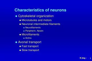
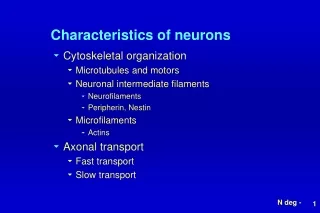
Toward Cryopreservation of Cultured Neurons. Hypertonic Solution. Isotonic Solution. Hypotonic Solution. Acrylic top cover. Rachel Bywater , Jenna Wilson, and Robert Zarfas School of Chemical, Biological, and Environmental Engineering. Acrylic gasket. Glass channel cover. Acrylic body.

E N D
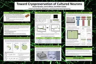
Toward Cryopreservation of Cultured Neurons Hypertonic Solution Isotonic Solution Hypotonic Solution Acrylic top cover Rachel Bywater, Jenna Wilson, and Robert Zarfas School of Chemical, Biological, and Environmental Engineering Acrylic gasket Glass channel cover Acrylic body Acrylic base Project objectives and goals Fluorescence quenching experiments Controlled-rate freezing experiments Enable long-term storage and stabilization of cultured neurons for use in cell-based biosensors - Determine permeability parameters for cultured neurons so that cryoprotectant chemicals (CPAs) can be added without cell death - Determine CPA concentrations and freezing rates which result in high cell viability Controlled-rate experiments are performed to determine optimal concentrations of CPAs and freezing rates that give high cell viability. A summary of the experiments to be performed in shown below. Permeability parameters can be determined for adherent cells through fluorescence quenching of calcein-AM, a fluorescent dye that can be transported into cells. The fluorescent intensity increases as the cell swells and decreases as the cell shrinks, giving a linear relationship between cell volume and intensity. During experimentation, the cells are exposed to solutions with different osmolarities and permeating characteristics using a flow chamber. The fluorescence intensity is recorded using a compound microscope with a green fluorescent filter and imaging software. During controlled rate freezing, cryoprotectant is first loaded into the cells. Next, the temperature is lowered until ice begins to form. At this point, the cells are frozen at a constant rate until they reach -80°C. The cells are then placed in liquid nitrogen for storage. What is cryopreservation? Why is it needed? When using permeating CPAs like propylene glycol, fluorescence quenching data looks like this: Due to the many advances in the areas of biotechnology and medicine, the need for long-term storage and stabilization of biological materials is rapidly increasing. Cryopreservation could potentially be used to maintain the integrity and functionality of proteins, cells, and organs while storing them outside of their native environment. Cryopreservation: - Cells and tissues are cooled to very low temperatures (-196°C), effectively stopping all biological activity Problems encountered in cryopreservation: - Solution effects/Cellular dehydration - Extracellular/Intracellular ice formation Cryoprotectants (CPAs) are chemicals used to prevent damage during freezing - Dimethylsulfoxide (DMSO), propylene glycol, and ethylene glycol are the most common CPAs CPA Addition Control Experiments Live/Dead Staining Technique The control experiments were performed to see how much cell damage was due only to cryoprotectant addition. This allows us to determine how much damage was due to the only the freezing process once all of the experiments have been performed. Live Stain Calcein-AM In order to determine how many cells survived addition of CPA and freezing, live and dead stains are applied. The mechanisms of each are shown. Dead stain: EthidiumHomodimer The data is adjusted for photobleaching of the fluorescent dye. The cell is initially exposed to an isotonic solution. When it is exposed to a hypertonic solution of propylene glycol, the cell shrinks. Because propylene glycol is able to permeate the cell membrane, the cell volume can equilibrate back to its initial volume. Summary of Permeability Parameters (at 4°C) Membrane permeability & osmotic pressure Green fluorescence (520 nm) Red fluorescence Osmotic pressure differences during addition and removal of CPAs can cause cell damage due to excessive swelling/shrinking. Determining the membrane permeability of the cells allows optimization of the addition and removal processes so that damage does not occur. The length of time necessary to load the cells with cryoprotectant is dependent on the specific chemical. This time is determined using the permeability parameters resulting from the fluorescence quenching experiments and the differential flux equations. Cells were loaded with cryoprotectant for the appropriate time. Damage was assessed both instantaneously and one day later. In all cases, approximately 5% of the cells were damaged due to addition of CPA. To 95% confidence, none of the experiments were determined to be significantly different. Green light (490 nm) Blue light The am groups are cleaved off by esterases inside live cells. Therefore, only live cells will contain fluorescent calcein. Ethidiumhomodimer can only penetrate “leaky” membranes. Therefore, only dead cells will contain the fluorescent dye. Flow chamber for fluorescence quenching A flow chamber is used in the fluorescent quenching experiments. The coverslip containing the adherent cells is placed face-down over a channel where solutions flow. The hole at the top of the chamber allows for the microscope objective to measure fluorescent intensity. The base acts as a heat exchanger so the experiments can be performed at various temperatures. Future Work The scheduled controlled-rate freezing experiments will be continued, and the cell viability will be determined for each condition. This will involve applying the live/dead stains and analyzing the cell viability. In addition, the permeability parameters determined in the fluorescence quenching experiments can be used to determine optimal methods of adding and removing cryoprotectants. This is done using a MatLab model, and the work will be performed by Allyson Fry. Differential Flux Equations: dVw/dt = volume flux of water across the cell membrane Πe/i= extracellular/intracellular osmotic pressure LP = hydraulic conductivity A = cellular surface area dVCPA/dt = molar flux of CPA across the membrane Me/i= extracellular/intracellular osmolarity PCPA = CPA permeability parameters A = cellular surface area CPA = CPA molar volume Acknowledgements We would like to thank the following people for their help and support: Dr. Adam Higgins (project sponsor), Dr. Phil Harding, Allyson Fry, and Manfred Dittrich