Download

1 / 78
780 likes | 873 Views
Explore the intricate world of cellular communication through chemical signals and signaling pathways, essential for both multicellular and unicellular organisms. Discover how cells respond to external signals, from receptor activation to cellular responses and apoptosis. Learn about the evolution of cell signaling and the role of signal transduction pathways in conveying messages within cells. Uncover the mechanisms behind local and long-distance signaling, including paracrine, synaptic, and endocrine signaling. Delve into the three stages of cell signaling: reception, transduction, and response, as elucidated by Earl W. Sutherland's groundbreaking research.

E N D
Chapter 11 Cell Communication
© 2011 Pearson Education, Inc. Overview: Cellular Messaging • Cell-to-cell communication is essential for both multicellular and unicellular organisms • Biologists have discovered some universal mechanisms of cellular regulation • Cells most often communicate with each other via chemical signals • For example, the fight-or-flight response is triggered by a signaling molecule called epinephrine
Cellular communication Contact Types of signaling Local Long-distance Paracrine Synaptic Neuronal: via elctricity Endocrine system: via hormones
Cellular communication Contact Types of signaling Local Long-distance Paracrine Synaptic Neuronal: via elctricity Endocrine system: via hormones
Cellular communication Contact Types of signaling Local Long-distance Paracrine Synaptic Neuronal: via elctricity Endocrine system: via hormones
Cellular communication Contact Types of signaling Local Long-distance Paracrine Synaptic Neuronal: via electricity Endocrine system: via hormones
Concept 11.1: External signals are converted to responses within the cell • Concept 11.2: Reception: A signaling molecule binds to a receptor protein, causing it to change shape • Concept 11.3: Transduction: Cascades of molecular interactions relay signals from receptors to target molecules in the cellConcept • 11.4: Response: Cell signaling leads to regulation of transcription or cytoplasmic activities • Concept 11.5: Apoptosis integrates multiple cell-signaling pathways
© 2011 Pearson Education, Inc. Concept 11.1: External signals are converted to responses within the cell • Microbes provide a glimpse of the role of cell signaling in the evolution of life
© 2011 Pearson Education, Inc. Evolution of Cell Signaling • The yeast, Saccharomyces cerevisiae, has two mating types, a and • Cells of different mating types locate each other via secreted factors specific to each type • A signal transduction pathway is a series of steps by which a signal on a cell’s surface is converted into a specific cellular response • Signal transduction pathways convert signals on a cell’s surface into cellular responses
1 2 3 factor Receptor How canthis beanalogized to people? Exchange of mating factors a a factor Yeast cell, mating type a Yeast cell, mating type Mating a New a/ cell a/
© 2011 Pearson Education, Inc. • Pathway similarities suggest that ancestral signaling molecules evolved in prokaryotes and were modified later in eukaryotes • The concentration of signaling molecules allows bacteria to sense local population density
1 2 3 Figure 11.3 Individualrod-shapedcells Aggregation in progress 0.5 mm Spore-formingstructure(fruiting body) 2.5 mm Fruiting bodies
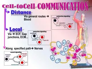
© 2011 Pearson Education, Inc. Local and Long-Distance Signaling • Cells in a multicellular organism communicate by chemical messengers • Animal and plant cells have cell junctions that directly connect the cytoplasm of adjacent cells • In local signaling, animal cells may communicate by direct contact, or cell-cell recognition
Plasma membranes Figure 11.4 Gap junctionsbetween animal cells Plasmodesmatabetween plant cells (a) Cell junctions (b) Cell-cell recognition
© 2011 Pearson Education, Inc. • Short-distance messenger molecules: local regulators • long-distance messenger molecules: hormones • The ability of a cell to respond to a signal depends on whether or not it has a receptor specific to that signal
Figure 11.5a Local signaling Electrical signalalong nerve celltriggers release ofneurotransmitter. Target cell Neurotransmitter diffuses across synapse. Secretingcell Secretoryvesicle Local regulatordiffuses throughextracellular fluid. Target cellis stimulated. (b) Synaptic signaling (a) Paracrine signaling
Long-distance signaling Figure 11.5b Endocrine cell Bloodvessel Hormone travelsin bloodstream. Target cellspecificallybinds hormone. (c) Endocrine (hormonal) signaling
© 2011 Pearson Education, Inc. The Three Stages of Cell Signaling: A Preview • Earl W. Sutherland discovered how the hormone epinephrine acts on cells • Sutherland suggested that cells receiving signals went through three processes • Reception • Transduction • Response
© 2011 Pearson Education, Inc. Animation: Overview of Cell Signaling Right-click slide / select “Play”
1 Figure 11.6-1 EXTRACELLULARFLUID CYTOPLASM Plasma membrane Reception Receptor Signalingmolecule
2 1 Figure 11.6-2 EXTRACELLULARFLUID CYTOPLASM Plasma membrane Reception Transduction Receptor Relay molecules in a signal transductionpathway Signalingmolecule
3 2 1 Figure 11.6-3 EXTRACELLULARFLUID CYTOPLASM Plasma membrane Response Reception Transduction Receptor Activationof cellularresponse Relay molecules in a signal transductionpathway Signalingmolecule
© 2011 Pearson Education, Inc. Concept 11.2: Reception: A signaling molecule binds to a receptor protein, causing it to change shape • The binding between a signal molecule (ligand) and receptor is highly specific • A shape change in a receptor is often the initial transduction of the signal • Most signal receptors are plasma membrane proteins
© 2011 Pearson Education, Inc. Receptors in the Plasma Membrane • There are three main types of membrane receptors • G protein-coupled receptors • Receptor tyrosine kinases • Ion channel receptors
G protein-coupled receptors (GPCRs) are the largest family of cell-surface receptorsThe G protein acts as an on/off switch: If GDP is bound to the G protein, the G protein is inactive Signaling molecule binding site Segment thatinteracts with G proteins G protein-coupled receptor
1 2 4 3 Note the enzyme is activated by shape change Figure 11.7b G protein-coupledreceptor Plasmamembrane Activatedreceptor Signalingmolecule Inactiveenzyme GTP GDP GDP CYTOPLASM Enzyme G protein(inactive) GTP GDP Activatedenzyme GTP GDP P i Cellular response
Figure 11.8: The sturcutre of a G Protien-Coupled Receptor Moleculeresemblingligand 2-adrenergicreceptors Plasmamembrane Cholesterol
© 2011 Pearson Education, Inc. • Receptor tyrosine kinases (RTKs) are membrane receptors that attach phosphates to tyrosines • Benefit: A receptor tyrosine kinase can trigger multiple signal transduction pathways at once • Tidbit: Abnormal functioning of RTKs is associated with many types of cancers
2 1 3 4 Signalingmolecule (ligand) Ligand-binding site Figure 11.7c Signalingmolecule helix in themembrane Tyr Tyr Tyr Tyr Tyr Tyrosines Tyr Tyr Tyr Tyr Tyr Tyr Tyr Tyr Tyr Tyr Tyr Tyr Tyr CYTOPLASM Receptor tyrosinekinase proteins(inactive monomers) Dimer Activated relayproteins Cellularresponse 1 P Tyr P Tyr P Tyr Tyr Tyr Tyr P Tyr P Tyr P P Tyr Tyr Tyr Tyr P Cellularresponse 2 Tyr P Tyr P Tyr Tyr P Tyr Tyr P 6 ADP 6 ATP Fully activatedreceptor tyrosinekinase(phosphorylateddimer) Activated tyrosinekinase regions(unphosphorylateddimer) Inactiverelay proteins
2 1 3 A ligand-gated ion channel receptor acts as a gate when the receptor changes shape When a ligand binds to the receptor, the gate allows specific ions, such as Na+ or Ca2+, through a channel in the receptor Figure 11.7d Gate closed Gate closed Ions Gate open Signalingmolecule (ligand) Plasmamembrane Ligand-gatedion channel receptor Cellularresponse
© 2011 Pearson Education, Inc. Intracellular Receptors • Intracellular receptor proteins are found in the cytosol or nucleus of target cells • Small or hydrophobic chemical messengers can readily cross the membrane and activate receptors • Examples of hydrophobic messengers are the steroid and thyroid hormones of animals • Outcome: An activated hormone-receptor complex can act as a transcription factor, turning on specific genes
EXTRACELLULARFLUID Hormone(testosterone) Figure 11.9-1 Plasmamembrane Receptorprotein DNA NUCLEUS CYTOPLASM
EXTRACELLULARFLUID Hormone(testosterone) Figure 11.9-2 Plasmamembrane Receptorprotein Hormone-receptorcomplex DNA NUCLEUS CYTOPLASM
EXTRACELLULARFLUID Hormone(testosterone) Figure 11.9-3 Plasmamembrane Receptorprotein Hormone-receptorcomplex DNA NUCLEUS CYTOPLASM
EXTRACELLULARFLUID Hormone(testosterone) Figure 11.9-4 Plasmamembrane Receptorprotein Hormone-receptorcomplex DNA mRNA NUCLEUS CYTOPLASM
EXTRACELLULARFLUID Hormone(testosterone) Figure 11.9-5 Plasmamembrane Receptorprotein Hormone-receptorcomplex DNA mRNA NUCLEUS New protein CYTOPLASM
© 2011 Pearson Education, Inc. Concept 11.3: Transduction: Cascades of molecular interactions relay signals from receptors to target molecules in the cell • Signal transduction usually involves multiple steps • What are some benefits of a multistep pathway?
© 2011 Pearson Education, Inc. Concept 11.3: Transduction: Cascades of molecular interactions relay signals from receptors to target molecules in the cell • Signal transduction usually involves multiple steps • Benefit: can amplify a signal: (A few molecules can produce a large cellular response) • Benefit: provide more opportunities for coordination and regulation of the cellular response
© 2011 Pearson Education, Inc. How Signal Transduction Pathways Work: • The molecules that relay a signal from receptor to response are mostly proteins • Like falling dominoes, the receptor activates another protein, which activates another, and so on, until the protein producing the response is activated • At each step, the signal is transduced into a different form, usually a shape change in a protein
© 2011 Pearson Education, Inc. Protein Phosphorylation and Dephosphorylation • In many pathways, the signal is transmitted by a cascade of protein phosphorylations • Protein kinases transfer phosphates from ATP to protein, a process called phosphorylation • Protein phosphatases remove the phosphates from proteins, a process called dephosphorylation • This phosphorylation and dephosphorylation system acts as a molecular switch, turning activities on and off or up or down, as required
Signaling molecule Figure 11.10 “upstream”/”downstream” regulation Receptor Activated relaymolecule Inactiveprotein kinase1 Activeprotein kinase1 Inactiveprotein kinase2 ATP Phosphorylation cascade ADP P Activeprotein kinase2 PP P i Inactiveprotein kinase3 ATP ADP P Activeprotein kinase3 PP P i Inactiveprotein ATP P ADP Activeprotein Cellularresponse PP P i
© 2011 Pearson Education, Inc. Small Molecules and Ions as Second Messengers • The extracellular signal molecule (ligand) that binds to the receptor is a pathway’s “first messenger” • Second messengers are small, nonprotein, water-soluble molecules or ions that spread throughout a cell by diffusion • Second messengers participate in pathways initiated by GPCRs and RTKs • Cyclic AMP and calcium ions are common second messengers
Figure 11.11 What other organic molecule do these resemble? Adenylyl cyclase Phosphodiesterase Pyrophosphate H2O P i P ATP cAMP AMP Why?
First messenger(signaling moleculesuch as epinephrine) Figure 11.12 Adenylylcyclase G protein GTP G protein-coupledreceptor ATP Second messenger cAMP Proteinkinase A Cellular responses
© 2011 Pearson Education, Inc. Calcium Ions and Inositol Triphosphate (IP3) • Calcium ions (Ca2+) act as a second messenger in many pathways • Calcium is an important second messenger because cells can regulate its concentration
EXTRACELLULARFLUID Plasmamembrane Figure 11.13 Ca2pump ATP Mitochondrion Nucleus CYTOSOL Ca2pump Endoplasmicreticulum(ER) Ca2pump ATP Low [Ca2 ] High [Ca2 ] Key