Download
1 / 33
330 likes | 369 Views
Learn how NMR spectroscopy aids in characterizing complex oligosaccharide structures. Discover how coupling constants, chemical shifts, and NOEs provide valuable insights. Explore techniques like COSY, ROESY, and NOESY to determine linkages and geometries. Understand how NOEs offer structural information, and use saturation transfer difference spectroscopy to identify ligand-protein interactions and bound geometry. See how angular constraints reveal molecular orientation for accurate structural analysis.

E N D
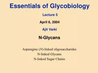
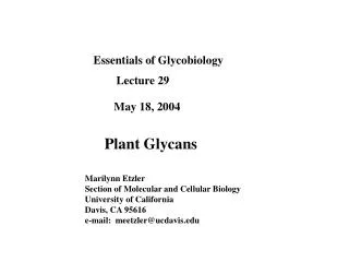
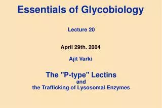
Glycobiology – BCMB 8130 NMR and Oligosaccharide Conformational Analysis
Useful References on NMR of Carbohydrates • 1. Prestegard, J.H., Bougault, C.M., and Kishore, A.I. (2004). Residual dipolar couplings in structure determination of biomolecules. Chemical Reviews 104, 3519-3540. • 2. Kogelberg, H., Solis, D., and Jimenez-Barbero, J. (2003). New structural insights into carbohydrate-protein interactions from NMR spectroscopy. Current Opinion in Structural Biology 13, 646-653. • 3. Bendiak, B., Fang, T.T., and Jones, D.N.M. (2002). An effective strategy for structural elucidation of oligosaccharides through NMR spectroscopy combined with peracetylation using doubly C-13-labeled acetyl groups. Canadian Journal of Chemistry-Revue Canadienne De Chimie 80, 1032-1050. • 4. Mayer, M., and Meyer, B. (2001). Group epitope mapping by saturation transfer difference NMR to identify segments of a ligand in direct contact with a protein receptor. Journal of the American Chemical Society 123, 6108-6117. • 5. Duus, J.O., Gotfredsen, C.H., and Bock, K. (2000). Carbohydrate structural determination by NMR spectroscopy: Modern methods and limitations. Chemical Reviews 100, 4589-+.
Glc II Glc T Carbohydrate Processing Enzymes in N-glycoside Biosynthesis Endoplasmic Reticulum Golgi Complex Bi-antennary ComplexI GlcNAc T V GnTI GnTII Golgi Man II Manstat Sw Golgi Man IA/IB ER Man I Glc II Glc I P OST P Tri-antennary Complex with b1,6-branch and polylactosamine Dol ER membrane Legend a1,2-Glc a1,2-Man a1,3-Glc a1,3-Man b1,2-GlcNAc a1,6-Man b1,4-Gal b1,4-Man a2,6-NeuNAc b1,4-GlcNAc b1,3-GlcNAc
Oligosaccharides are Structurally Very Diverse • Consider six common sugar residues, Glc, GlcNAc, Gal, Man, NeuNAc, Fuc • Consider 4 possible linkage sites • Consider α and β linkages • Consider branched in addition to linear • There are 64x43x24 + 64x42x24x3 primary structures for a tetrasaccharide • Or: 2,322,432 structures
Oligosaccharide Characterization from 1D 1H Spectra • Anomeric resonances are easily identified and counted • Anomeric configuration correlates with coupling constants and chemical shift • Acetylation, methylation easily identified • Residue identification and linkage requires more complex spectra
MicrocoilNMR Probesoffer High SensitivityforLigandScreeningand Characterization
Two Dimensional NMR SpectraA General Scheme: other mixing and evolution periods can be added to increase dimensions Preparation Evolution 1 (Increment t1) Mixing Evolution 2 (observe t2) time Example: COSY – mixing is scalar coupling 90x 90x (mix) d1 (recover) t1 (evolve) t2 (observe)
Expansion of Sialyl Lewis X Ring Region:Galactose Assignment
z z z z z y y y y y x x x x x 90x 90x 90x mix d1 (recover) t1 (evolve) t2 (observe) 2D NOE Spectroscopy (NOESY) a b b a Some magnetization precessing at a in t1 can precess at b in t2
Periodic Inversion Sets Stage for Magnetization Transfer During Mixing Time r 1H 1H M For short T, Large c, I 1/r6 T = 0 I T = T
NOE (Nuclear Overhauser Effect)depends on competition between W0 and W2 processes (0) ( /2) irradiate W2 () (3/2) () (/2) W0 (2) (3/2) (/2) relax, assuming W0 dominates observe E (3/2- ) (/2+) (3/2)
NOEs are Positive for Small Molecules, Negative for Large = (-1 + 6/(1+402c2))/(1 + 3/(1+02c2) + 6/(1+402c2)) 1/2 1.1/ 0 0 c -1
In Practice Data May be Collected for Cross Peaks at a Series of Mixing Times • Icp = C{exp(-T) • (1 – exp(-2T)} • = 2W1 + W2 + W0 , = (W2 - W0) dIcp/dT 1/r6 Icp T
r12 r14’ NOEs Give Structural Information 14’ / 12 = r126 / r14’6 r12 = 2.5 Å, 14’ / 12 = 4.0 , implies: r14’ = 3.15 Å
GnT-Vlarge, glycosylated, expresses only in mammalian cells, no experimental structure, but can we investigate bound ligand geometry? ? ?
Saturation transfer difference spectroscopy can identify primary areas of contact Mayer M & Meyer B: JACS, 123:6108-6117(2001))
Saturation transfer difference spectra indicate the UDP portion to be most tightly associated
Transfer NOEs Accentuate the Bound State And Give Geometry Information Protein sipb K-1 sijf sijb K1 sjpb Ligand *Cross-relaxation rate c, r, 0 *Chemical exchange fast w.r.t cross-relaxation rate and chemical shift scale *Observed NOE is weighted average of free and bound states *Change in sign of NOE upon change in molecular weight
Sign Change of Cross-Peak Confirms Binding Solution Geometry Retained UDP-GlcNAc UDP-GlcNAc and GnT-V
Ligand Geometry from Orientational Data 3 1H 15N
Dipole-Dipole Interactions Give Angular ConstraintsIndependent of Distance B0 1H q r • distance dependence (fixed by bonding) • angular dependence (long range information) 13C However, when molecules tumble isotropically in solution - all orientations sample equally, - <3Cos2q-1> averages to zero
Introducing Molecular Order B0 Anisotropic case: - some orientations are preferred over others - dipolar coupling is directly observable - a variety of media exist phage, bicelles, C12E5/hexanol, etc
Couplings Can be Easily Measured in Coupled HSQC Spectra
RDC Measurements for UDP and Acceptor with GnTV Natural abundance Coupled 1H-13C HSQC spectrum C B A 291.7 293.7 294.6 UH5’ UH2’ 150.4 150.0 148.6 AH1 151.3 152.1 150.2 GnT-V impurity A] UDP (6mM) + Acceptor (4mM) only B] UDP (6mM) + Acceptor (4mM) in 10 mg/ml phage C] UDP (6mM) + Acceptor (4mM) + GnT-V (0.2 mM) in 10 mg/mL phage
Principal Alignment Frames for Bound State Ligands of GnT-V Sxx Sxx Szz Szz Syy Syy Order Tensor for UDP Order Tensor for Acceptor • Both ligands are bound to the protein and experience a common ordering due to the protein • The molecular frames of the ligands can be rotated until their order tensors match => Gives relative geometry of the ligands
Orientations of Donor and Acceptor in GnT-V Binding Site from RDC Analysis