Download
1 / 97
970 likes | 981 Views
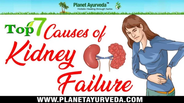
This article discusses the manifestations of renal failure, including integumentary changes, electrolyte imbalances, alterations in acid/base balance, cardiovascular and gastrointestinal problems, endocrine issues, and anemia. It provides management strategies for these manifestations.

E N D
ManifestationsofRenal FailureAlice Hellebrand MSN, RN, CNN, CURNMay 7, 2011ANNA Jersey North 126
Manifestations of Renal Failure • Alterations in integument • Electrolyte imbalance • Alterations in acid/base balance • Alterations in cardiovascular system • Alterations in gastrointestinal system • Endocrine problems • Anemia
Alterations in Integument • Signs/Symptoms: Skin that is grayish-bronze; pale, dry, and scaly skin, pruritis, ecchymosis • Etiology: Retained urinary pigments, anemia, decreased activity of sweat and sebaceous glands, uremic toxins and calcium phosphate deposits in skin, sensory nerve irritation, capillary fragility, abnormal platelet adhesiveness
Alterations in Integument • Management: ~Moisturize skin with super-fatted soaps, bath oils, and lotions ~Anti-pruritic medications ~Correct calcium/phosphate imbalances with meds and dialysis ~Dialysis
Regulates water and fluid balance Can cause high blood pressure by holding onto extra water Hypernatremia – Excessive sodium can cause tissue swelling (edema) Hypernatremia can cause the water in the cells to exit = crenation Hyponatremia – Too little sodium causes water to move into the cells = hemolysis Electrolyte ImbalanceSodium (Na+)
Involved in nerve and muscle function, contraction of the heart muscle Hyperkalemia – Too much potassium can cause the heart to beat irregularly or even stop Signs and Symptoms 1. Muscle weakness 2. Tall-tented T-waves 3. Feel your heart beating (irregular) 4. Cardiac arrest Hypokalemia – Too little potassium Extreme muscle weakness; hard to walk Potassium (K+)
Most of the calcium is within bone and teeth Regulates blood clotting Regulates enzymes Regulates hormone action Controls function of nerves and muscles Hypercalcemia – Confusion, lethargy, loss of appetite, nausea/vomiting, and abdominal pain Hypocalcemia – Seizures, tetany, numbness, increase PTH Calcium (Ca++)
Hyperphosphatemia – Severe itching; crystal deposition under the skin, in blood vessel walls, and in the heart muscle Lowers the levels of calcium, causing increase PTH excretion Hypophosphatemia – Weakness, coma, and bone softening Phosphate (PO4-)-phosphorus
Helps regulate acid-base balance (pH) Normal kidneys regenerate and keep bicarbonate; failed kidneys cannot Bicarbonate is a base, therefore it neutralizes acid Too little bicarbonate = metabolic acidosis Too much bicarbonate = metabolic alkalosis Bicarbonate – (HCO3-)
Metabolic Acidosis • Etiology: ~Inability of kidneys to excrete hydrogen ions ~Reduction in ammonia synthesis in renal tubular cells = decreased excretion of ammonium chloride ~Inability of kidneys to reabsorb bicarbonate ions to buffer excess acids ~Decreased ability of kidneys to produce and excrete titratable acids (HPO4/H2PO4) ~Retention of acid end products of metabolism ~Catabolism of body proteins
Metabolic Acidosis • Oral alkaline medications (bicarbonate) • Intravenous sodium bicarbonate • Dialysis • Correct catabolism
Cardiovascular System • Hyperkalemia • Hypertension • Pericarditis
Etiology GFR = K+ secretion Metabolic acidosis Catabolism of body proteins Bleeding Blood transfusions Dietary indiscretion Meds and IVs with K+ Management: Monitor intake Correct catabolism Avoid salt substitutes Correct acid/base Dialysis Pharmacologic therapy ~Cation exchange, hypertonic glucose and insulin, IV bicarb Hyperkalemia
Etiology Excess fluid and sodium contributing to vascular volume overload Malfunction of the renin-angiotensin (R-A) system Management Control fluid and salt intake Dialysis to remove fluid and sodium Antihypertensives ACE inhibitors to control R-A system Hypertension
Etiology Inflammation of pericardial membrane due to uremic toxins, occasionally bacterial and viral infections Chest pain, fever, friction rub Can progress to effusion and/or tamponade Management Daily dialysis for two weeks without heparin Anti-inflammatory to reduce the swelling Pericarditis
Signs and Symptoms Oral: Fetor uremicus, gum ulcers, bleeding, metallic taste, stomatitis Anorexia, nausea and vomiting Stomach and intestines: Gastritis w/bleeding, gastric/duodenal ulcers, constipation/diarrhea Management Frequent oral care Dialysis Bulk-forming laxatives or stool softeners Antidiarrheals Gastrointestinal System
Children: Stop growing ~Growth hormone, anemia, abnormal protein metabolism, acidosis, Ca/PO4 imbalance Failure to menarche Failure to develop secondary sex characteristics Adults: Females: Decreased libido, failure to ovulate, amenorrhea, abnormal hormone levels Males: Decreased libido, impotence, decreased sperm production, testicular atrophy, low testosterone levels Endocrine Function
Etiology Shortened life span GI bleeding Blood loss during dialysis Decreased red blood cell production Infection and inflammation Hemolysis Iron deficiency Management Iron supplementation Folic acid supplement Correct uremia EPO Blood transfusion Androgen therapy Anemia
Gross Renal Anatomy • Kidneys – Retroperitoneal • Weight - 120-160 gm • Size - 2x4 inches by 1 inch thick • Nephron is the functional unit • Ureters – Peristalsis • Bladder – Storage tank • Urethra – Exit from body Source: American Nephrology Nurses’ Association, 1993.
Internal Structure of the Kidney • Cortex - 85% cortical nephrons; 15% juxtamedullary nephrons • Blood flow faster through cortex than medulla • Medulla – Pyramids, renal columns, loops of Henle, vasa recta • Pyramids - Contain nephrons and their blood vessels • Collecting ducts pelvis Source: American Nephrology Nurses’ Association, 1993.
Renal Blood Supply • Receives ~25% of cardiac output • Blood enters via afferent arteriole • Glomerulus filters plasma • Blood exits via efferent arteriole • Peritubular capillary network - All cortical, most of juxtamedullary • Vasa recta – Juxtamedullary nephron loops of Henle only Source: American Nephrology Nurses’ Association, 1993.
Tubular Component • Bowman’s capsule – Houses the glomerulus • Proximal convoluted tubule • Loop of Henle • Distal convoluted tubule • Collecting ducts Source: American Nephrology Nurses’ Association, 1993.
Juxtaglomerular Apparatus • Decreased blood pressure sensed by macula densa cells in the afferent arteriole stimulates the juxtaglomerulus apparatus to secrete renin • It is converted to Angiotensin I in the liver, then converted to Angiotensin II in lung tissue • Vasoconstriction and sodium and water retained = BP Source: American Nephrology Nurses’ Association, 1993.
Nephron Functions: Water and Electrolyte Regulation • Tubular reabsorption • Tubular secretion • Clearance
Water and Electrolyte Balance • Glomerulus • Plasma filtered into Bowman’s capsule • RBCs, WBCs, and proteins are not filtered • Sympathetic innervation in response to decreased blood flow (bleeding, hypotension): • Afferent arteriole vasoconstricts • Glomerulus permeability decreased
Water and Electrolyte Balance • Proximal Tubule • 65% of Na+ actively reabsorbed • Chloride andH2O follow passively • 100% glucose and amino acids reabsorbed • Most K+ reabsorbed • Some Mg++, Ca++ and PO4- reabsorbed • Acid-base balance begins • Filtrate leaves isotonic
Water and Electrolyte Balance • Loop of Henle • Counter current multiplying and exchange mechanism established between long, thin loops of Henle of juxtamedullary nephrons and adjacent vasa recta • Ascending limb has diluting mechanisms: Filtrate leaves hypotonic
Water and Electrolyte Balance • Distal Tubule • Sodium and potassium are regulated by aldosterone • Water is reabsorbed with sodium
Water and Electrolyte Balance • Collecting Tubule • Sodium and potassium regulated by aldosterone • Antidiuretic hormone (ADH) regulates water reabsorption (by making distal tubule permeable to water) and determines final urine concentration and volume
Water and Electrolyte Balance • ADH - “Antidiuretic Hormone” • Same as “vasopressin” • Released by pituitary gland • Triggered by osmoreceptors in the hypothalamus and baroreceptors in the aortic arch • Makes distal tubules permeable to water • Adjusts osmolarity
Other Renal Functions: • RBC production regulated by erythropoetin secretion • Secreted in response to renal hypoxia • Acts on bone marrow to increase the rate of RBC production • Metabolism of Vitamin D
Acute Renal Failure • Sudden rapid deterioration • Severe • Most common causes • Hypoperfusion • Nephrotoxins • Often reversible
Acute Renal Failure • Mortality rate – 40-60% • Etiology – Characterized by location and cause • Pre-renal • Intra-renal • Post-renal
Acute Renal Failure • Sudden, severe, often reversible • Goal: Prevent life-threatening complications, such as infection and GI bleeding • Remove the cause, restore kidney function
Acute Renal Failure • Pre-Renal – Before the kidney • Intra-Renal – Within the kidney • Post-Renal – After the kidney Source: American Nephrology Nurses’ Association, 1993.
Pre-renal Causes • Hypovolemia • Altered peripheral vascular resistance • Cardiac disorders
Intra-renal Causes • Nephrotoxic Agents • Drugs • Contrast media • Biological substances • Environmental agents • Heavy metals • Plant and animal substances
Intra-renal Causes • Inflammatory processes • Trauma • Radiation nephritis • Obstruction • Intravascular hemolysis • Systemic and vascular disorders
Post-renal Causes • Obstruction • Ureteral • Bladder neck • Uretheral • Prostatic hypertrophy • Abdominal or pelvic neoplasms • Pregnancy • Neurogenic bladder
Chronic Renal Failure • Insidious, progressive, irreversible • 1995 – 257,000 people with ESRD • 2002 – 300,000+ people with ESRD • Causes: • #1 Diabetes • #2 Hypertension
Other Causes • Glomerulonephritis • Polycystic kidney disease • Interstitial nephritis • Obstructions – Birth defects, blocking objects, and scarring
Other Causes (cont) • Drug toxicity - Heroin and other recreational drugs - Nonsteroidal anti-inflammatories (NSAIDS) - Antibiotics - Anti-rejection medications
Principles of Dialysis • FLOW – Determined by the amount of blood delivered by the heart (pump). • RESISTANCE – Factors that slow down the flow of blood throughthe blood vessels. • PRESSURE – Combination of flow from the heart and resistance in the blood vessels.
Dialysate • A solution containing the electrolytes (calcium, sodium, bicarbonate, potassium, magnesium, and chloride) in the same concentration as the blood stream of people with normal kidney function.
Diffusion • The movement of particles from an area of higher solute concentration to an area of lower concentration via a semipermeable membrane. Source: American Nephrology Nurses’ Association, 1993.
Osmosis • The movement of fluid (water) from an area of lower solute concentration to an area of higher solute concentration. Source: American Nephrology Nurses’ Association, 1993.
Ultrafiltration • The movement of fluid with additional pressure applied, either positive or negative via a semipermeable membrane.