Understanding Renal Failure: Causes, Symptoms, and Management
Explore the structure and function of the kidney, different types of renal failure, key diagnostic tests, assessment of symptoms, and nursing interventions for acute and chronic renal failure. Learn about risk factors, key factors, and management strategies.

Understanding Renal Failure: Causes, Symptoms, and Management
E N D
Presentation Transcript
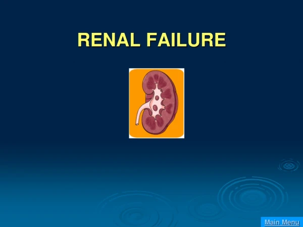
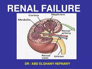
Structure and Function of the Kidney • Primary unit of the kidney is the nephron • 1 million nephrons per kidney • Composed of a glomerulus and a tubule • Kidneys receive 20% of cardiac output Renal Lecture Required Picture #1
Key points • The kidneys “ • Regulate fluid, • Regulate acid-base, • Regulate electrolyte balance, and • Eliminatingwastes from the body. • ????? • RF may be acute /chronic • ARF: sudden interruption of renal function. (obstruction, poor circulation, or kidney disease.
Acute Renal Failure - Definitions • Reversible • 70% Non-oliguric , 30% Oliguric • Non-oliguric associated with better prognosis and outcome • “Overall, the critical issue is maintenance of adequate urine output and prevention of further renal injury.”
ARF • ARF is comprised of three phases: • Oliguria – begins with the renal insult and continues for 3 weeks. • Diuresis – begins when the kidneys begin to recover and continues for 7 to 14 days. • Recovery – continues until renal function is fully restored and requires 3 to 12 months. • Prerenal failure from hemorrhage or prolonged hypotension is the most common cause of acute renal deterioration and is usually reversible with prompt intervention.
Chronic Renal Failure (CRF) • a progressive, irreversible kidney disease. • End-stage renal failure exists when 90% of the functioning nephrons have been destroyed and are no longer able to maintain fluid, electrolyte, or acid-base homeostasis.
Key Factors • Risk factors for ARF may be classified as: • Prerenal, (hypovolemia, decreased cardiac output, Decreased peripheral vascular resistance, renal vascular obstruction) • Intrarenal, (Nephrotoxic injury, Acute glomerulonephritis) or • Postrenal. bilateral obstruction of urine outflow (???)
Key Factors • Risk factors of CRF: • ARF. • DM HTN • Chronic glomerulonephritis. • Nephrotoxic medications (????) or chemicals. • Pyelonephrosis • Autoimmune disorders (SLE). • Polycystic kidney. • Renal artery stenosis recurrent UTI
Renal Failure - Diagnosis • Urinalysis • Hematuria, proteinuria, and alterations in specific gravity • Serum creatinine: gradual increase of 1 to 2 mg/dL per every 24 to 48 hr for ARF • Gradual increase over months to years for CRF • Blood urea nitrogen (BUN)
Renal Failure - Diagnosis • Serum electrolytes • Dilutionalhyponatremia & hypocalcemia • Increased potassium, phosphorus, and magnesium • CBC; ???? • Ultrasound • KUB • CT
Renal Failure - Diagnosis • Aortorenal angiography • Cystoscopy • Retrograde pyelography • Renal biopsy • Nuclear medicine scans
Assessment • S&Sx occur suddenly with ARF. • Client with CRF may be asymptomatic except during periods of stress (infection, surgery, and trauma). • In most cases, symptoms are related to fluid volume overload and include: • Renal – polyuria, nocturia (early), oliguria, anuria (late), proteinuria, hematuria, and dilute urine color when present.
Assessment • Cardiovascular – HTN, peripheral edema, pericardial effusion, CHF, cardiomyopathy, and orthostatic hypotension. • Respiratory – dyspnea, tachypnea, uremic pneumonitis, lung crackles, Kussmaul respirations, and pulmonary edema. • Hematologic – anemia, bruising, and bleeding.
Assessment • Neurologic – lethargy, insomnia, confusion, encephalopathy, seizures, . • GI – A, N, V, metallic taste, stomatitis, diarrhea, uremic halitosis. • Skin – decreased skin turgor, yellow cast to skin, dry, pruritus, and bruising • osteomalacia, muscle weakness, pathologic fractures, and muscle cramps. • Reproductive – erectile dysfunction.
Assessment • Report: • Urinary elimination patterns (amount, color, odor, and consistency). • Vital signs (especially blood pressure). • Weight – 1 kg (2.2 lb) daily weight increase is approximately 1 L of fluid retained. • Assess/monitor vascular access or peritoneal dialysis insertion site.
NANDA Nursing Diagnoses • Imbalanced nutrition • Risk for infection • Impaired gas exchange • Activity intolerance • Impaired skin integrity • Disturbed thought processes • Deficient knowledge
ARF - Management • Nutrition management • Initially very catabolic • Goals: • Adequate calories • Low protein • Low K and Phos • Decreased fluid intake
Nursing Interventions • Provide high carbohydrate and moderate fat content in the client’s diet. • Restrict the client’s intake of fluids (based on urinary output). • Balance the client’s activity and rest. • Prepare the client for hemodialysis.
Nursing Interventions • Provide skin care to prevent breakdown. • Protect the client from injury. • Provide emotional support to the client and family. • Encourage the client to ask questions • Encourage the client to diet, exercise, and take medication to control hyperlipidemia.
Nursing Interventions • Administer medications as prescribed. • Antihypertensives– • Iron supplements and folic acid as needed • Erythropoietin • Vitamin D supplements and calcium supplements • Stool softeners • Diuretics (except in ESRD)
For clients with ARF, the nurse should: • Identify and assist with correcting the underlying cause. • Prevent prolonged episodes of hypotension and hypovolemia. • Prepare for fluid challenge and diuretics Restrict fluid intake , Na, & K during oliguric phase.
For clients with CRF, the nurse should: • Obtain a detailed medication Hxto determine the client’s risk • Control protein intake • Restrict the client’s dietary Na, K, ph, and Mg • Refer the client to a community resource or support group.
Client with CRF • Encourage the client to stop smoking . • Encourage diabetic client to adhere to strict blood glucose control ???? • Teach the client how to measure BP & Wt at home.
Complications and Nursing Implications • Hyperkalemia – Administer Kayexalate or insulin as prescribed. • HTN – Administer antihypertensives and diuretics as prescribed. • Seizures – Implement seizure precautions. • Cardiac dysrhythmias – Provide life support interventions for lifethreatening • dysrhythmias. Monitor the client for and report non-lethal dysrhythmias.
Complications and Nursing Implications • Pulmonary edema – Prepare the client for hemodialysis. • Infection – Maintain the client’s surgical asepsis of invasive lines. Monitor the client for signs of localized and systemic infections and report. • Metabolic acidosis – Prepare the client for hemodialysis. • Uremia – Prepare client for hemodialysis.s
Renal Replacement Therapy • Peritoneal Dialysis • Acute Intermittent Hemodialysis • Continuous Hemofiltration
Function of Dialysis • Rid the body of excess fluid & electrolytes. • Achieve acid-base balance. • Eliminate waste products. • Restore internal homeostasis • Dialysis can sustain life. • Dialysis does not replace the hormonal functions of the kidney.
Hemodialysis • Shunts the client’s blood from the body through a dialyzer and back into the client’s circulation. • Requires internal or external access device.
Therapeutic Procedures and Nursing Interventions • Prior to hemodialysis, assess for patency of the access site (presence of bruit, palpable thrill, distal pulses, and circulation). Before and after assess: Vital signs. • Laboratory values (BUN, serum creatinine, electrolytes, hematocrit). • Weight.
After hemodialysis assess: • For complications (hypotension, access clotting, headache, muscle cramps, hepatitis). • Access site for indications of bleeding, infection. • For nausea, vomiting, level of consciousness.. (hypovolemia)
Nursing Interventions • Discuss with Dr any medications to be withheld until after dialysis. Provide emotional support prior • Avoid taking BP, administering injections, performing venipuncturesor inserting IV lines on an arm with an access site. • Avoid invasive procedures (4 - 6 hr) after d • Elevate the extremity following surgical development of AV fistula to avoid swelling.
Teach the client to • Avoid lifting heavy objects with access-site arm. • Avoid carrying objects that compress the extremity. • Avoid sleeping with body weight on top of the extremity with the access device. • Perform hand exercises that promote fistula maturation.
Complications and Nursing Implications • Hemodialysis • Clotting/Infection of Access Site • Use sterile technique • Avoid compression of access site/extremity • Hypotension • Discontinue dialysis. • Place the client in the Trendelenburg position. • Anemia: Administer prescribed medication Infectious Diseases espbloodbornediseasess • .