ECG’s
230 likes | 576 Views
ECG’s. Jake Turner. What is an ECG?. A recording of the electrical activity within the heart. What you need to know . Basic pathologies that can be picked up on ECG How to calculate heart rate Shockable rhythms How to localise a pathology from an ECG Basic arrhythmias. ECG basics.

ECG’s
E N D
Presentation Transcript
ECG’s Jake Turner
What is an ECG? • A recording of the electrical activity within the heart.
What you need to know • Basic pathologies that can be picked up on ECG • How to calculate heart rate • Shockable rhythms • How to localise a pathology from an ECG • Basic arrhythmias
ECG basics • Check that this ECG is for the patient in front of you! (Name, DOB, patient number etc) • Check which lead the rhythm strip is (usually lead II) • At the bottom left is the 'paper speed' (25 mm/s on the horizontal axis) and the sensitivity of the ECG (10mm/mV).
How to calculate heart rate • Method 1: We always print off 10 second ECG strips, so count the number of QRS complexes, multiply this by 6 and you have the heart rate! • Method 2: Count the number of large squares between each QRS complex, then divide 300 by this number (this method cannot be used for an irregular rhythm). • NOTE: To calculate the heart rate using method 1 you must use the rhythm strip!
Arrhythmias on ECG • Ventricular or atrial • Too fast, too slow or irregular • Sinus rhythm, regularly regular • Normal, tachycardic or bradycardic • Sinus rhythm, regularly irregular • P-P interval varies by more than 10%. • Irregularly irregular • Atrial fibrillation (VF is effectively pulseless)
Sinus rhythm • This just means that every QRS complex is preceded by a P wave! • Note: It does not necessarily mean that every P wave is followed by a QRS complex.
How to tell if a rhythm is regular? • Check if the ECG printout tells you! • Paper strip method
How to read an ECG (the official version) • Step 1: Rhythm • Step 2: Rate • Step 3: Conduction (PQ,QRS,QT) • Step 4: Heart axis • Step 5: P wave morphology • Step 6: QRS morphology • Step 7: ST morphology • Step 7+1: Compare the current ECG with a previous one
What we need to read from an ECG • Step 1: What jumps out at you? (VF, VT, irregularly irregular, gross morphological problems, ST elevation indicative of an NSTEMI etc) • Step 2: Rhythm • Step 3: Rate • Step 4: Conduction (is there conduction?) • Step 5: General morphology (is everything about the right size?) • Step 6: Compare the current ECG with a previous one (this is less likely to come up in an OSCE, but could do in an exam)
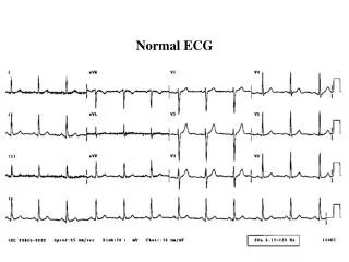
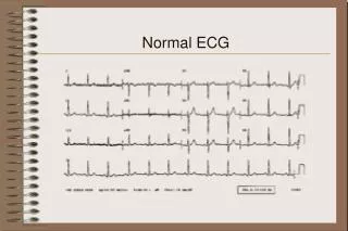
Normal ECG • Rhythm: sinus • Rate: 60-100 bpm • PQ interval 120-200ms • QRS width 60-100ms • Heart axis: between -30 and +90 degrees • The maximal height of the P wave is 2.5 mm in leads II and / or III • The p wave is positive in II and AVF, and biphasic in V1 • The p wave duration is usually shorter than 0.12 seconds (3 small squares) • No pathological Q waves • No left or right ventricular hypertrophy • Normal R wave propagation. (R waves increase in amplitude from V1-V5) • No ST elevation or depression • T waves should be concordant with the QRS complex • The ECG should not have changed from the previous ECG
What didn’t I cover? • Heart blocks • The effects of ion disturbances • QRS complex abnormalities • Bundle branch blocks • Cardiac hypertrophy • Genetic conditions • Treatments • Axis deviation
ECG denotations. • The letters "Q", "R" and "S" are used to describe the QRS complex • Q: the first negative deflection after the p-wave. If the first deflection is not negative, the Q is absent. • R: the positive deflection • S: the negative deflection after the R-wave • Small print letters (q, r, s) are used to describe deflections of small amplitude. For example: qRS = small q, tall R, deep S. • R`: is used to describe a second R-wave.
Revision sites • http://en.ecgpedia.org/wiki/Main_Page • http://en.ecgpedia.org/wiki/Basics • http://www.medicine-on-line.com/html/ecg/e0001en_files/14.htm • http://www.nottingham.ac.uk/nursing/practice/resources/cardiology/index.php
Specific revision pages • For bundle branch blocks: http://www.medicine-on-line.com/html/ecg/e0001en_files/13.htm • For right and left hypertrophy: http://www.medicine-on-line.com/html/ecg/e0001en_files/12.htm