Download

1 / 47
500 likes | 1.1k Views
THE ECG. Dr. George John Critical Care, Christian Medical College, Vellore. Reading the ECG. Rate Rhythm Axis Chamber Enlargement Myocardial Damage Miscellaneous References: http://library.med.utah.edu/kw/ecg/ecg_outline/Lesson1/index.html. ECG – BASIC NOMENCLATURE. Rate. Axis.

E N D
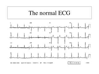
THE ECG Dr. George John Critical Care, Christian Medical College, Vellore
Reading the ECG Rate Rhythm Axis Chamber Enlargement Myocardial Damage Miscellaneous References: http://library.med.utah.edu/kw/ecg/ecg_outline/Lesson1/index.html
Chamber Enlargement The ECG criteria for diagnosing right or left ventricular hypertrophy are very insensitive (i.e., sensitivity ~50%, which means that ~50% of patients with ventricular hypertrophy cannot be recognized by ECG criteria). However, the criteria are very specific (i.e., specificity >90%, which means if the criteria are met, it is very likely that ventricular hypertrophy is present).
LVH - 1 • S in V1 + R in V5 or V6 > 35 mm • R in aVL >11 mm or, if left axis deviation, R in aVL >13 mm plus S in III >15 mm • CORNELL Voltage Criteria for LVH (sensitivity = 22%, specificity = 95%) S in V3 + R in aVL > 24 mm (men) S in V3 + R in aVL > 20 mm (women)
LVH - 2 ESTES Criteria for LVH ("diagnostic", >5 points; "probable", 4 points)
RVH • V1 Lead: - R/S ratio > 1 and negative T wave - R > 6 mm, or S < 2mm, - rSR' with R' >10 mm • R in V1 + S in V5 (or V6) > 10 mm • V5 or V6 - R/S ratio in V5 or V6 < 1 - R in V5 or V6 < 5 mm - S in V5 or V6 > 7 mm
LAESensitivity = 50%; Specificity = 90% • P wave duration > 0.12s in frontal plane (usually lead II) • Terminal P negativity in lead V1 (i.e., "P-terminal force") duration >0.04s, depth >1 mm.
RAE • P wave amplitude >2.5 mm in II and/or >1.5 mm in V1 (Sensitivity = 50%; Specificity = 90%) • QRS voltage in V1 is <5 mm and V2/V1 voltage ratio is >6 (Sensitivity = 50%; Specificity = 90%) Criteria derived from the QRS complex are due to both the high incidence of RVH when RAE is present, and the RV displacement by an enlarged right atrium.
Myocardial Damage • Type of damage: Change in ECG • Location: Leads involved
Myocardial DamageLocation Limb Leads: L2, aVF, L3: Inferior L1, aVL: Lateral aVR: Cavity Chest Leads: V1, V2: Anterior V3, V4: Septal V5, V6: Lateral
Infarct / Necrosis • Q Wave • Non Q Wave Pathologic Q waves are usually defined as duration >0.04 s or >25% of R-wave amplitude)
Q Wave Infarcts Pathologic Q waves, ST elevation, T wave inversion (acute necrosis) Pathologic Q waves, T wave inversion (necrosis and fibrosis) Pathologic Q waves, upright T waves (fibrosis)
Non Q MI Two-thirds of MI's presenting to emergency rooms evolve to non Q wave MI's, most having ST segment depression or T wave inversion. Recognized by evolving ST-T changes over time without the formation of pathologic Q waves (in a patient with typical chest pain symptoms and/or elevation in myocardial-specific enzymes)
ST - T Changes 1 PRIMARY ST-T Changes: Intrinsic myocardial disease (e.g., myocarditis, ischemia, infarction, infiltrative or myopathic processes) Drugs (e.g., digoxin, quinidine, tricyclics, and many others) Electrolyte abnormalities of potassium, magnesium, calcium Neurogenic factors (e.g., stroke, hemorrhage, trauma, tumor, etc.) Metabolic factors (e.g., hypoglycemia, hyperventilation)
ST-T Changes 2 "SECONDARY" ST-T Wave changes: ST-T changes seen in bundle branch blocks (generally the ST-T polarity is opposite to the major or terminal deflection of the QRS) ST-T changes seen in fascicular block ST-T changes seen in nonspecific IVCD ST-T changes seen in WPW preexcitation ST-T changes in PVCs, ventricular arrhythmias, and ventricular paced beats
T & U Waves • The T wave is the most labile wave in the ECG • The U wave is the only remaining enigma of the ECG The normal U wave has the same polarity as the T wave and is usually less than one-third the amplitude of the T wave.
ELECTROCARDIOGRAMS Dr. George John, Critical Care, Christian Medical College, Vellore
ECG - K 70 YEAR OLD MAN WITH GIDDINESS:
THE END THANK YOU!! Acknowledgement: http://www.ecglibrary.com/ Dean Jenkins and Stephen Gerred.