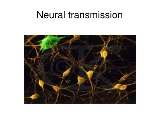
Neural Transmission
Neural Transmission. Glial cells and moving across the synapse. Again: Three Steps for firing. Resting potential : voltage is about -70mV Dendrites receive incoming signals If sufficient, cell goes into firing mode Action potential Voltage changes from -70mV to +40mV

Neural Transmission
E N D
Presentation Transcript
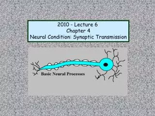
Neural Transmission Glial cells and moving across the synapse
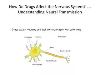
Again: Three Steps for firing • Resting potential: voltage is about -70mV • Dendrites receive incoming signals • If sufficient, cell goes into firing mode • Action potential • Voltage changes from -70mV to +40mV • Ions exchange places • Repeats itself rapidly down axon • Only in places where myelin sheath doesn’t cover: Nodes of Ranvier • Refractory Period: • below resting or lower than -70mV • Cell recovers from firing • Absolute refractor period: Brief time period when cannot fire again • Relative refractory period: Brief time period when difficult for it to fire again. • Take home lesson: • Axon encodes stimulus intensity by controlling FIRING RATE not size of action potential
The Neuron Fires • Action potential causes nearby Na+ channels to open, so another action potential is triggered right next to first one, and this continues all the way down the axon • Chain reaction • Like a bunch of dominoes • Action potential ≠ local potential in several important ways: • Local potential = graded potential- it varies in magnitude depending on strength of stimulus that produced it; action potential is ungraded • Action potential obeys all or none law: occurs at full strength or not at all • Action potential is nondecremental: does NOT lose strength at each successive point (local potentials do degrade)
Why so fast?Thank your Glial cells • Review: Glial cells: • Non neural cells • Provide a supporting function to neurons • Account for 90% of cells in adult human brain • Function: Help hold neurons together, assist in neurotransmission • Provide supports for the nervous system • In periphery are rather rigid: • E.g., Schwann cells • In CNS: are soft and squishy: • E.g., Oligodenroglia cells
Glial Cells speed conduction • Neurons conduct impulses from 1 to 120 meters/sec or 270 mph • Still fairly slow • Since reaction time critical for survival, body found ways to increase conduction speed • Vary thickness of axons to provide less resistance • Motor neurons: diameter of 0.5 mm can attain conduction speed of 30m/sec • But: conduction speed not increase in direct proportion to size: is power function, thus must find alternative way • Alternative way: use graded local potentials
Myelination is solution to increasing speed of neural transmission • Vertebrates are myelinated • Glial cells produce myelin • Fatty tissue that wraps around axon • Insulates it from surrounding interstitial fluid • Forms what look like beads on a string • Gaps are open to interstitial fluid: NODES OF RANVIER • Membrane exposed • Sodium channels here • Graded potentials can trigger action potential • Thus: in vertebrates, SALTATORY conduction • Action potential jumps from node to node • Myelin also helps increase speed via capacitance: resists movement of ions during graded potential • Overall effect: 100x greater conduction speed; reduced work for Ion pump
Other functions of Glial Cells • Important in fetal development: scaffold that guides new neurons to destination • Provide energy to cell • Serve as waste system for neurons • Aid in development and maintenance of neural connections • Get 7x more connections when glial cells available • Help in conducting action potential
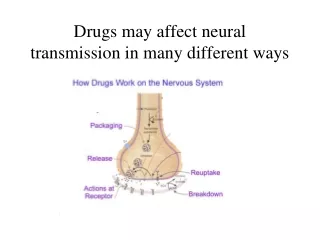
Now: Why an action potential? • Allows release of neurotransmitter • Neurotransmitter is chemical • Several specific kinds- each act on certain neurons • Most neurons respond to and release one kind of neurotransmitter • Neurotransmitter stored in synaptic vesicles • Action potential opens channels that allow Ca+ ions to enter terminals from extracellular fluid • Ca+ ions cause vesicles nearest the membrane to fuse with membrane • Membrane then opens and transmitter is dumped into synapse • Diffuses across synapse to postsynaptic neuron and attaches to chemical receptor
Action in the Synapse • Neurotransmitter is released into the synapse • diffuses across synapse to next neuron’s dendrite • This “next dendrite” is post-synaptic • Neurotransmitter is attracted to the POST-synaptic side: • receptor sites on the next neurons dendrites • attach if can find right spot: neurotransmitter must match molecular shape of receptor site • Activation of receptor causes ion channels in membrane to open • Ionotropic receptors open channels directly to produce immediate reactions required for motor and sensory processing • Metabotropic receptors open channels indirectly and more slowly to produce longer-lasting effects • Sets off graded potentials for next action potential • Movement across the synapse is relatively slow: several milliseconds
Excitation and Inhibition • The NT opens ion channels on dendrites and soma • Two effects on local membrane potential: • shifts in positive direction (towards 0), partially depolarizing • Shifts in negative direction (away from 0): hyperpolarization • Thus two effects: • Excitatory: depolarization • Inhibitory: hyperpolarization
Two kinds of postsynaptic potentials: • EPSPs: excitatory postsynaptic potentials • Excitatory effect: increases likelihood of action potential • Opens Na+ channels • IPSPs: inhibitory postsynaptic potentials • Inhibitory effect: decreases likelihood of action potential • Opens K+ channels • Thus: bidirectional effects • Summative effects • Overall change must be sufficient to produce action potential
Postsynaptic integration • Summation across all the IPSPs and EPSPs • Summates algebraically • Adds both positive and negatives together • Two kinds: • Spatial summation: • Sum of all IPSPs and EPSPs occurring simultaneously at different locations along dendrites and cell body • Must be sufficient number of “hits” • Temporal summation • Sum of all IPSPs and EPSPs occurring within a short time • Must occur within a few milliseconds • Must get sufficient number of “hits” within certain time • Neuron is an information integrator! • A decision maker • Small microprocessor
Terminating synaptic activity • Neurons are efficient • Not all neurotransmitter is attached to post-synaptic receptor sites • Extra must be destroyed or repackaged • Enzymes in synapse attack and destroy extra NT • e.g., Monoamine oxidase or MAO, acetylcholinesterase • Attach and degradate neurotransmitter • MAO inhibitors stop this process and prolong action of dopamine, norepinephrine and serotonin in synapse: • e.g., Elavil • Reuptake • Neuron takes NT back up and recycles it for later use • There is a specialized autoreceptor that detects how much extra • Specialized transporter attaches to NT and brings it back into cell • SSRI, NSRI and SNSRI drugs do this block this action • E.g., prozac, lexapro, wellbutrin, etc.
So: Put it all together • Neuron receives incoming NT which attaches to receptor sites on dendrites • Results in local potentials: excitatory or inhibitory • If sufficient, produce an action potential • The action potential involves exchange of ions and opening of cell wall along axon hillock and nodes of Ranvier • The action potential is nondecremental or saltatory conduction • Results in moving down of synaptic vessicles and fusing of synaptic vessicles to terminal button wall • Neurotransmitter is released in synapse • Most makes it to the next neuron (dendrites) • Some may be degraded by enzymes • Some may be reuptaken • Some may float away • And so it all begins again, billions of times per minutes.