Download
1 / 52
520 likes | 665 Views
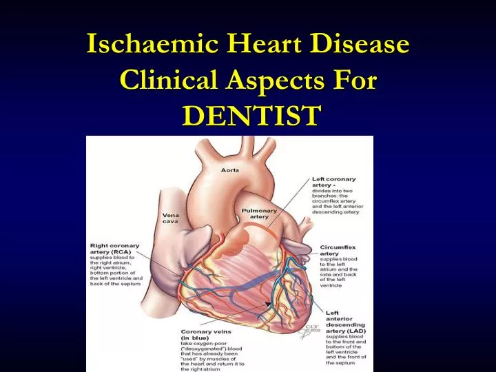
Ischaemic Heart Disease Clinical Aspects For DENTIST. Coronary Artery Disease. A leading cause of SICKNESS and DEATH. Coronary Heart Disease: Myocardial Ischemia. An imbalance between the supply of oxygen and the myocardial demand resulting in myocardial ischaemia .

E N D
Coronary Artery Disease A leading cause of SICKNESS and DEATH
Coronary Heart Disease: Myocardial Ischemia • An imbalance between thesupplyofoxygenand themyocardialdemandresulting in myocardial ischaemia. • Decreased blood supply (and thus oxygen) to the myocardium that can result in acute coronary syndromes: • Angina pectoris ( Stable ) • Unstable Angina • Myocardial infarction • Sudden death (due to fatal arrhythmias)
Coronary Artery Diseases Coronary artery disease :Is the presence of atherosclerosis in the coronary arteries. Angina pectoris : Is a transient discomfort (usually less than 15 minutes) due to a temporary lack of adequate blood supply to the heart muscle. (symptom not a disease ) due to increased demands (Anaemia, hypertension, high cardiac output (thyrotoxicosis, myocardial hypertrophy) Acute Coronary Syndrome: Unstable Angina & Myocardial infarction Due to coronary vessel obstruction
Acute Coronary Syndromes Unstable Angina Angina that is continuing, prolonged or occurring at rest. Represents a syndrome that lies between angina pectoris and AMI. Myocardialinfarction (Heart Attack)is defined as death of heart tissue due to blockage of a coronary artery caused by atherosclerosis and thrombus formation
Ischaemic heart diseaseEpidemiology • Commonest cause of death in the Western world. (up to 35% of total mortality) • Over 20% males under 60 years have IHD • Health Survey : 3% of adults suffer from angina 1% have had a myocardial infarction in the past 12 months
Risk Factors for Cardiovascular Disease • Hypertension • High cholesterol • Obesity • Cigarette smoking • Physical inactivity • Diabetes mellitus • Kidney disease • Older age (>55 ♂; > 65 ♀) • Family history of premature cardiovascular disease • Periodontal disease ?
Ischaemic heart diseaseAetiology • Fixed • Age, Male, +ve family history • Modifiable – strong association • Dyslipidaemia, smoking, diabetes mellitus, obesity, hypertension • Modifiable - weak association • Lack of exercise, high alcohol consumption, type A personality, Oral Contraceptive Pills, soft water • PRIMARY PREVENTION Atherosclerosis
Ischaemic heart diseaseManifestations • Sudden death • Acute coronary syndrome ( Myocardial Infarction & Unstable Angina ) • Stable angina pectoris • Heart failure • Arrhythmia • Asymptomatic
Angina Pectoris • At least 70% occlusion of coronary artery resulting in pain. • Chest pain • - Characteristics: squeezing, bursting, pressing, burning or choking - Location: substernum - Refer pain: L’t shoulder, arm, neck or mandible - Associated with exertion, anxiety - Relieved by vasodilator (ex. NTG) or rest - May accompanied by dyspnea, nausea& vomiting sensation, palpitation • Usually brought on by physical exertion • Is self limiting usually stops when exertion is ceased
Angina pectoris Potential problem related to dental care 1. Stress and anxiety related to dental visit may precipitate angina attack Prevention of complication 1. Detection of patient 2. Referral of patient for medical evaluation and treatment 3. Known case with medical treatment for angina Stress reduction protocol Premedication Open and honest communication Morning appointments Short appointments Nitrous oxide - oxygen Avoid excessive amounts of epinephrine
Terminate all procedures • Semi-reclined position • Sublingual NTG • O2 • Check vital signs Discomfort relieved Still discomfort after 3min 6. Assume angina pectoris was present 7. Slowly taper O2 over 5min 8. Modify dental treatment Give 2nd NTG Still discomfort after 3min Give 3rd NTG Still discomfort after 3min • Angina Pectoris NTG 0.6mg/tab
10. Assume myocardial infarction in progress 11. On IV line 12. Prepare transport to ER If highly suspected AMI MONA:Morphine, Oxygen, NTG, Aspirin
Myocardial Infarction • Partial or total occlusion of one or more of the coronary arteries due to an atheroma, thrombus or emboli resulting in cell death (infarction) of the heart muscle • When an MI occurs, there is usually involvement of 3 or 4 occluded coronary vessels
Chest Pain • Site Jaw to navel, retrosternal, left submammary • Radiation Left chest, left arm, jaw….mandible, teeth, palate • Quality/severity tightness, heaviness, compression…clenched fists • Precipitating/relieving factors physical exertion, cold windy weather, emotion rest, sublingual nitrates • Autonomic symptoms sweating, pallor, peripheral vasoconstriction, nausea and vomiting
Differential diagnosis • Cardiacpathology • Pericarditis, aortic dissection • Pulmonarypathology • Pulmonary embolus, pneumothorax, pneumonia • Gastrointestinalpathology • Peptic ulcer disease, reflux, pancreatitis, ‘café coronary’ • Musculoskeletalpathology • Trauma, Tietze’s Syndrome
Assessment • 30% of deaths occur in the first 2 hours. (Cardiac muscle death occurs after 45 minites of ischaemia) • Symptoms and signs of myocardial ischaemia • Also • Changes in heart rate /rhythm • Changes in blood pressure
Confirming the diagnosis • Typical chest pain • Electrocardiographic changes ( ECG ) • ST elevation • new LBBB • Myocardial enzyme elevation • Creatine kinase (CK-MB) • Troponin
Treatment • Stop dental treatment • Call for help • Rest, sit up and reassure patient • Oxygen • Analgesia (opiate, sublingual nitrate) • Aspirin • Thrombolysis • Primary angioplasty • Beta-Blockers • ACE inhibitors • Prepare for basic life support • Transport patient to hospital
Surgical Treatment • Percutaneous Transluminal Coronary Angioplasty (PTCA) • balloon expansion that can provide 90% dilitation of vessel lumen • Stent Placement • Coronary Artery By-Pass Graft (CABG)
Acute Myocardial InfarctionComplications • Sudden Death (18% within 1 hour,36% within 24 hours) • Non-fatal arrhythmia • Acute left ventricular failure • Cardiogenic shock • Papillary muscle rupture and mitral regurgitation • Myocardial rupture and tamponade • Ventricular aneurysm and thrombus • Distal Embolisation
Prevention of complication 1. No routine dental care until at least 6 months after infarction 2. Medical consultation Current status Medication used 3. Stress reduction protocol Premedication Open and honest communication Morning appointment Short appointment Nitrous oxide - oxygen 4. Avoid excessive amounts of epinephrine 5. Check PT (Anticoagulant medication)
Dental Considerations for IHD • Common Situations: • Orthostatic Hypotension due to use of anti-hypertensives (beta blockers, nitroglycerin…) • Raise chair slowly • Allow patient to take his/her time • Assist patient in standing • Post-Op Bleeding: • When patients on Plavix or Aspirin, expect increased bleeding because of decreased platelet aggregation • Emergent Situations: • Possible MI: • Remember that pain in the jaw may be referred pain from the myocardium assess the situation, have good patient history, follow ABC’s • Angina: • In situations of angina pectoris, all operatories should have nitroglycerin to be placed sublingually
RISK FOR DENTAL PROCEDURE • Major Risk for Perioperative Procedures: • Unstable Angina (getting worse) • Recent MI • Intermediate Risk for Perioperative Procedures: • Stable Angina • History of MI • Most dental procedures, even surgical procedures fall within the risk of less than 1% • Some procedures fall within an intermediate risk of less than 5% • Highest risk procedures those done under general anesthesia
Dentistry & Cardiovascular Medicine • AMI • GA within 3/12 of AMI: 30% re-infarction rate @ 1/52 post op • Avoid routine LA dental treatment for 3/12 (emergency treatment only) • Avoid excess dosage, reduce anxiety • Avoid elective surgery under GA for1 year (specialist) • Be aware of medications (bleeding, hypotension)
Post MI: When to Treat • Why delay treatment? • Remember that with an MI there is damage to the heart, be it severe or minimal that may effect the patient’s daily life • MI within 1 month Major Cardiac Risk • MI within longer then 1 month: • Stable routine dental care ok • Unstable treat as Major Cardiac Risk • Older studies suggest high re-infarction rates when surgery performed within 3 months, 3-6 months… however, this was abdominal and thoracic surgery under general anesthesia • New research suggests delaying elective tx for 1 month is advisable. Emergent care should be done with local anesthetic without epinephrine and monitoring of vital signs • When in doubt: • CONSULT THE CARDIOLOGIST
Dental Management: Stable Angina/Post-MI >4-6 weeks • Minimize time in waiting room • Short, morning appointments • Preop, intra-op, and post-op vital signs • Pre-medication as needed • anxiolytic (triazolam; oxazepam); night before and 1 hour before • Have nitroglycerin available – may consider using prophylacticaly • Use pulse oximeter to assure good breathing and oxygenation • Oxygen intraoperatively (if needed) • Excellent local anesthesia - use epinephrine, if needed, in limited amount (max 0.04mg) or levonordefrin (max. 0.20mg) • Avoid epinephrine in retraction cord
Dental Management:Unstable Angina or MI < 3 months • Avoid elective care • For urgent care: be as conservative as possible; do only what must be done (e.g. infection control, pain management) • Consultation with physician to help manage • Consider treating in outpatient hospital facility or refer to hospital dentistry • ECG, pulse oximetry, IV line • Use vasoconstrictors cautiously if needed
Intraoperative Chest Pain • Stop procedure • Give nitroglycerin • If after 5 minutes pain still present, give another nitroglycerin • If after 5 more minutes pain still present, give another nitroglycerin • If pain persists, assume MI in progress and activate the EMS • Give aspirin tablet to chew and swallow • Monitor vital signs, administer oxygen, and be prepared toprovide life support
Warning Signs and Symptoms of Heart attack • Pressure, fullness or a squeezing pain in the center of chest that lasts for more than a few minutes. • Pain extending beyond the chest to the shoulder, arm, back or even your teeth and jaw. • Increasing episodes of chest pain • Prolonged pain in the upper abdomen • Shortness of breath- may occur with or without chest discomfort • Sweating • Impending sense of doom • Lightheadedness • Fainting • Nausea and vomiting
Emergency action plan for a person with signals of heart attack Unknown case of CAD Recognize the signals of a heart attack Stop activity and sit or lie down Wait about 5 minutes to see if the symptom go away. If the pain persists : Known case of CAD Recognize the signals of heart attack Stop activity and sit or lie down Take 1 nitroglycerin tablet at a time at 3 - to 5 minutes intervals to maximum total dose of 3 tablets. If pain persists. Transport patient to hospital
Conclusion: • When treating patients with Ischemic Heart Disease or recent MI… • Use caution and common sense • When in doubt: • CONSULT THE CARDIOLOGIST
What is Heart Failure? • HF occurs when the heart is unable to pump enough blood to meet the oxygen requirements of the body • Nearly 10% of populations > 70 years of age will have HF • Overall mortality close to 20% • HF risk factors: • CAD and its sequelae • HTN - Myocarditis, • Cardiomyopathy - Valvular heart diseases • Pericardial disease - Pulmonary embolism
Types and Classifications of H.F(measured by ejection fraction [EF]) • Systolic or diastolic • High output or low output • Left or right sided • Acute or chronic
Systolic HF Diastolic HF • Inability of heart to contract strongly enough to provide adequate blood flow to periphery • Abnormal relaxation of myocardium resulting in reduced filling of ventricle Systolic dysfunction: - EF < 50%; results from reduced left ventricular function - Increased preload - Most cases of CHF
Common Causes of HF Coronary Heart Disease/MI Hypertension Valvular Heart Disease Arrhythmias Myocarditis Cardiomyopathy Infective Endocarditis Congenital Heart Disease Pulmonary Hypertension Endocrine Disorders (thyroid disease) MI is a leading cause
Right Heart Failure Systemic venous congestion (distended neck veins, enlarged liver, peripheral edema, ascites) Left Heart Failure Pulmonary edema (Dyspnea) Sequelae of Heart Failure
Symptoms of Heart Failure Compensated (Asymptomatic) Uncompensated (Symptomatic) • Fatigue • Dyspnea • Orthopnea • Paroxysmal Nocturnal Dyspnea • Ankle Edema • Weight Gain Note: patients with a very low EF may have no symptoms
Functional Classification of Heart Failure (NYHA) • Class I: No limitation of physical activity. No dyspnea, fatigue, or palpitations with ordinary physical activity • Class II: Slight limitation of physical activity. Fatigue, palpitations and dyspnea with ordinary physical activity but comfortable at rest. • Class III: Marked limitation of activity. Less than ordinary physical activity results in symptoms but comfortable at rest. • Class IV: Symptoms present at rest and any physical activity exacerbates the symptoms
Medical Management of Heart Failure • Decreased cardiac output CO • Decreased ejection fraction • - repair of diseased valves • Fluid overload • Overweight • HTN Main Problems requiring treatment
Medical Management of Heart Failure • Treatment of underlying disease • Life-style modifications • Drug therapy • ACE inhibitors - Or angiotensin receptor blockers • Beta Blockers (Coreg, Toprol-XL, or bisoprolol) • Diuretics - Or direct-acting vasodilators • Nitrates • Digitalis Glycosides • Heart transplant
Dental Management Considerations (Heart Failure) • For undiagnosed pt with symptoms of HF: avoid elective care; refer to physician • For patients with diagnosed HF: • Class I (asymptomatic): routine care • Class II (mild symptoms with exertion): elective care OK and recommend consultation with physician • Class III or IV (symptoms with minimal activity or at rest): avoid elective care; if treatment necessary, manage in consultation with physician; consider referral to a special patient care setting; avoid use of vasoconstrictors