Download
1 / 55
550 likes | 655 Views
Learn the mechanism of radiation damage, exposure and toxicity assessment processes, data collection, evaluation, and risk characterization. Understand the phases of physical, chemical, and biological effects of ionizing radiation on DNA and cells.

E N D
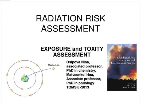
RADIATION RISK ASSESSMENT Osipova Nina, associated professor, PhD in chemistry, Matveenko Irina, Associate professor, PhD in philology TOMSK -2013 EXPOSURE and TOXITY ASSESSMENT
The contents • 1.What is the mechanism of radiation damage? • 2.Exposure assessment • 3. TOXICITY ASSESSMENT
the four basic steps in the risk assessment process: • 1. Data Collection and Evaluation • 2. Exposure Assessment • 3. Toxicity Assessment • 4. Risk Characterization
ExposureAssessment • is the process of estimating or measuring the magnitude, frequency and duration of exposure to an agent, alongwith the number and characteristics of the population exposed
ACRONYMS, SYMBOLS, AND UNITS • A(t) = Activity at Time t • Bq = Becquerel • Ci = Curie • D = Absorbed Dose • DCF = Dose Conversion Factor Per Unit Intake • HE = Effective Dose Equivalent HT = Dose Equivalent Averaged Over Tissue or Organ T HE,50 = Committed Effective Dose Equivalent Per Intake HT,50 = Committed Dose Equivalent Averaged Over Tissue T • LET = Linear Energy Transfer • LLD = Lower Limit of Detection • MeV = Million Electron Volts pCi = PicoCurie (10-12 Ci) • Q = Quality Factor in Definition of Dose Equivalent • RBE = Relative Biological Effectiveness • SI = International System of Units • Sv = Sievert • T = Tissue or Target Organs • wT = Weighting Factor in the Definition of Effective Dose Equivalent and Committed Effective Dose Equivalent
The absorption of energy from ionizing radiation produces damage to molecules by direct and indirect actions.For direct action, damage occurs as a result of ionization of atoms on key molecules in the biologic system. This causes inactivation or functional alteration of the molecule.Indirect action involves the production of reactive free radiacals whose toxic damage on the key molecule results in a biologic effect.
A free radical is an electrically neutral atom with an unshared electron in the orbital position. The radical is highly reactive.
Sequence of Effects • Physical: less than seconds • Chemical: seconds • Biological: seconds to many years • Reactions with molecules and cells • Tissue changes • Cancer, leukemia
Physical phase • Interactions of radiation with matter • Direct ionization of atoms by charged particles • Indirect ionization by neutral particles
Chemical phase • Chemical changes of biological molecules • The rates of chemical effects are comparable with the rates of chemical reactions.
Biological phase • A damaged cell may die. • A damaged cell may be mutated.
The transfer of the free radical to a biologic molecule can be sufficiently damaging to cause bond breakage or inactivation of key functions. The organic peroxy free radical can transfer the radical form molecule to molecule causing damage at each encounter. Thus a cumulative effect can occur, greater than a single ionization or broken bond. DNA is the primary target for cell damage from ionizing radiation. Toxic effects at low doses to moderate doses (cell killing, mutagenesis, and malignant transformation) appear to result from damage to cellular DNA. Thus, ionizing radiation is a classical genotoxic agent.
DNA is the most important material making up the chromosomes and serves as the master blueprint for the cell. It determines what types of protein that are produced in cell.
The DNA molecule takes the form of a double helix. The sides of the chain are strands of alternating sugar and phosphate groups. Branching off from each sugar group is one of four nitrogenous bases: cytosine, thymine, adenine and guanine. This large molecule is sensitive to radiation damage.
Ionising radiation can induce a wide variety of DNA lesions: breaks, base changes or cross-links, among others
Radiation emitted by radioactive substances can transfer sufficient localized energy to atoms to remove electrons from the electric field of their nucleus (ionization). In living tissue, this energy transfer can produce chemically reactive ions or free radicals, destroy cellular constituents, and damage DNA. Irreparable DNA damage is thought to be a major factor in carcinogenesis. • While ionizing radiation may also cause other detrimental health impacts, only radiogenic cancer risk is normally considered in risk assessments
low linear energy transfer (LET) radiation (photons and electrons) and high-LET radiations (alpha particles and neutrons) The type of ionizing radiation emitted by a particular radionuclide depends upon the exact nature of the nuclear transformation, and may include emission of alpha particles, beta particles (electrons or positrons), and neutrons; each of these transformations may be accompanied by emission of photons (gamma radiation or x-rays). Each type of radiation differs in its physical characteristics and in its ability to inflict damage to biological tissue. For purposes of radiation risk estimates, the various types of radiation are often categorized as low linear energy transfer (LET) radiation (photons and electrons) and high-LET radiations (alpha particles and neutrons)
Low dose exposures • Low dose exposures are not clinically detectable. That means that the long term effects can only be • Estimated in statistical studies of whole populations. In this way, the estimated effects are compared to other • common societal health risks (driving, flying, sports, alcohol, etc.) to look for the smallest discernible • increased risk. This might mean that an increase of one additional case of cancer in a population of a million • would be noted
low exposure levels • Because of the inability to observe definite effects of low exposure levels in living tissue, the most con-servative approach is used when evaluating risks from radiation exposure. This approach is called the Linear No-Threshold Risk Model which assumes some minimal biological effect from any exposure even though it would only apply statistically to large populations — not to individuals
The biological effects of low doses can be divided into three categories: • 1. Genetic Effects. Observed in offspring of exposed individuals resulting from egg or sperm cell muta- • tions. As in the case of chemically (drugs) or biologically (viruses) related mutations, these effects • usually result in nonviable organisms, but never in the “monsters” portrayed in movies or comics. • 2. Somatic Ef fects. Observed in the exposed individuals. These might take the form of cancers similar • to the effects of chemical agents (smoking or drinking alcohol) or biological agents (viruses). • 3. In-Utero E ffects. Observed after birth due to exposure of the embryo. This is a sub-set of somatic • effects, more serious in earlier stages of development, probably resulting in death, with diminishing • consequences later in fetal development.
Stochastic Effects • No threshold dose exist for these effects. • Can be somatic or hereditary (genetic). • Can occur after exposure to moderate-low doses of radiation. • These effects occur long-term after irradiation. • The probability of occurrence, but not the severity, increase with dose.
acute doses • In contrast to the low doses over long time, there have been occasions in which humans have received • large doses in a matter of hours. These are called acute doses . In the cases of the atomic bombing of Hiroshi- • ma and Nagasaki and the catastrophic release of radioactive material from the Chernobyl power plant in the • former Soviet Union, the clinical effects were detectable. Cells were killed with resulting organ and whole • body damage. Table 1 briefly describes the expected effects from acute doses.
absorbed dose • The average energy imparted by ionizing radiation per unit mass of tissue. The SI unit of absorbed dose is the joule per kilogram, also assigned the special name the Gray (1 Gy = 1 joule/kg); the conventional unit of absorbed dose is the rad • (1 rad = 0.0 1 Gy).
What are radionuclide slope factors? • slope factors are used for estimating incremental cancer risks resulting from exposure to radionuclides via inhalation, ingestion, and external exposure pathways.
Slope factors for radionuclides - the probability of cancer incidence as a result of unit exposure to a given radionuclide averaged over a lifetime. It is the age-averaged lifetime excess cancer incident rate per unit intake (or unit exposure for external exposure pathway) of a radionuclide
Dose conversion factors (DCFs), or "dose coefficients” • for a given radionuclide represent the dose equivalent per unit intake (i.e., ingestion or inhalation) or external exposure of that radionuclide. These DCFs are used to convert a radio- nuclide concentration in soil, air, water, or foodstuffs to a radiation dose. DCFs may be specified for specific body organs or tissues of interest, or as a weighted sum of individual organ dose, termed the effective dose equivalent
What is dose equivalent, effective dose equivalent, and related quantities • "dose equivalent“isa measure of the energy absorbed by living tissues, adjusted for the relative biological effectiveness of the type of radiation present.
dose equivalent dose equivalent = the absorbed dose X Quality Factor (Q) or radiation weighting factor
The "committed dose equivalent" • is defined as the integrated dose equivalent that will be received by an individual during a 50-year period (based on occupational exposure) following the intake.
absolute excess risks of death in a cohort of people with cancer • AER = ((O-E)/person-years) * 10,000 (the observed (O) and expected (E) deaths) This gives us the excess risk per year per 10,000 persons. We have done this for various age groups and would like to use a test for trend to see whether there is a significant increase in the risk
The "critical organ" is the organ that received the most dose for the radionuclide concerned • Critical organ standards • usually consist of a combination of whole body and critical • organ dose limits, such as • 25 mrem/yr to the whole body, • 75 mrem/yr to the thyroid, 25 mrem/yr to any critical organ other than the thyroid. When these standards were • adopted, dose was calculated and controlled for each organ • in the body and uniform radiation of the "whole body." • The "critical organ" was the organ that received the most • dose for the radionuclide concerned. With the adoption of • the dose equivalent concept, the dose to each organ is • weighted according to the effect of the radiation on the • overall system (person).
Distribution of polonium-210 in human body organs after chronic ingestion of radionuclides of uranium and thorium series
radionuclide slope factors are used to estimate the excess cancer risk resulting from exposure to radionuclides at radiologically contaminated sites for comparison with target risk range • 10-4-10-6 lifetime excess cancer risk
The incremental risk • is calculated by multiplying estimates of the lifetime intake via inhalation and ingestion of each radionuclide of concern, and the duration and concentration in environmental media to which the receptor is exposed via the external exposure pathway, by the appropriate slope factor values for that exposure pathway and radionuclide.
The incremental risk • lifetime intake x slope factor, • where slope factor takes account of the duration and concentration in environmental media to which the receptor is exposed
Regulatory Limits • Radiation worker: • 5 rem/yr = 50 mSv/yr • Individual member of the general public: • 0.1 rem/yr = 1 mSv/yr Compare to • natural background: 0.06 rem/yr = 0.6 mSv/yr • average background: 0.36 rem/yr = 3.6 mSv/yr • LD50/30: 200-300 rad = 2-3 Gy