BONE HISTOLOGY
450 likes | 634 Views
BONE HISTOLOGY. Dr. Nabil Khouri. Review: Hyaline Cartilage. Provides support, flexibility, and resilience Is the most abundant skeletal cartilage Is present in these cartilages: Articular – covers the ends of long bones Costal – connects the ribs to the sternum

BONE HISTOLOGY
E N D
Presentation Transcript
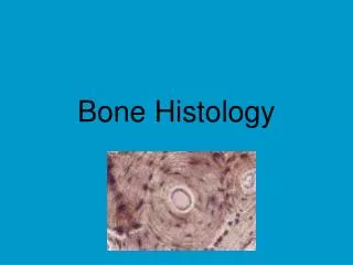
BONE HISTOLOGY Dr. Nabil Khouri
Review: Hyaline Cartilage • Provides support, flexibility, and resilience • Is the most abundant skeletal cartilage • Is present in these cartilages: • Articular – covers the ends of long bones • Costal – connects the ribs to the sternum • Respiratory – makes up the larynx and reinforces air passages • Nasal – supports the nose
HYALINE CARTILAGE A Thechondrocytesare located in Clacunae BThematrixcontain collagen fibers that are so fine they are not visible in tissue preparations.
Bone Anatomy • Diaphysis • Metaphysis • Epiphysis – Prox/Dist • Epiphyseal line • Periosteum • Compact cortical bone • Spongy bone • Articular Cartilage • Medullary cavity • Marrow • Nutrient artery
Histology of bone tissue Cells are surrounded by matrix. - 25% water - 25% protein - 50% mineral salts Cellulerity 4 cell types make up osseous tissue Osteoprogenitor Osteoblasts Osteocytes Osteoclasts
Osteogenic cells are the only bone cells that divide. Osteogenic cells differentiate and develop into osteoblasts which, in turn, are responsible for forming new bone. Osteoblasts synthesize and secrete a collagen matrix and calcium salts. When the area surrounding an osteoblast calcifies, the osteoblast becomes trapped and transforms into an osteocyte, the most common and mature type of bone cell. Osteoclasts, the cells that break down and reabsorb bone, stem from monocytes and macrophages rather than osteogenic cells. There is a continual balance between osteoblasts generating new bone and osteoclasts breaking down bone.
Osteo-progenitor cells: derived from mesenchyme bone connective tissue is derived from - unspecialized stem cells - Undergo mitosis and develop into Osteoblasts - found on inner surface of periosteum and endosteum.
Osteoblasts: • Are large cell responsible for the synthesis and mineralization of bone (bone forming cells) during both initial bone formation and later bone remodeling. • Found on surface of bone form a closely packed sheet - No ability to mitotically divide - Collagen secretors • They arise from the differentiation of osteogenic cells in the periosteum, the tissue that covers the outer surface of the bone, and in the endosteum of the marrow cavity. • The osteoblasts produce many cell products, including the enzymes alkaline phosphatase and collagenase, growth factors, hormones such as osteocalcin, and collagen, part of the organic unmineralized component of the bone called osteoid.
Osteocytes: Mature bone cells inside the bone derived form osteoblasts Do not secrete matrix material Cellular duties include: exchange of nutrients and waste with blood. Some of the osteoblasts turn into osteocytes while the new bone is being formed, and the osteocytes then get surrounded by new bone. They are not isolated, however, because they send out long branches that connect to the other osteocytes. These cells can sense pressures or cracks in the bone and help to direct where osteoclasts will dissolve the bone.
Osteoclasts Are bone resorping cells found in pits in the bone surface which are called resorption bays, or Howship's lacunae. Roll : growth, maintenance and bone repair • Osteoclasts are large multinucleate cells differentiate from another type of cell called a macrophage. • Osteoclasts are formed by the fusion of many cells derived from circulating monocytes in the blood.
Bone Tissue: Supportive Connective Tissue Extracellular Matrix 25% Water 25% Protein or organic matrix 95% Collagen Fibers 5% Chondroitin Sulfate 50% Crystalized Mineral Salts Hydroxyapatite (Calcium Phosphate) Other substances: Lead, Gold, Strontium, Plutonium, etc.
Two types of Bone tissue Compact Bone Spongy Bone
Compact Bone: External layer Compact & Spongy - lamellar bone Called lamellar bone (groups of elongated tubules called lamella) It is arranged in units called osteons (The Haversian systems). Osteons contain blood vessels, lymphatic, nerves - blood vessels and nerves penetrate periosteum through horizontal openings called perforating (Volkmann’s) canals. Surrounding this canal are concentric rings of osteocytes along with the calcified matrix.
- Osteons Osteon is concentric rings (lamellae) of calcified matrix surrounding a vertically oriented blood vessel are aligned in the same direction along lines of stress. These lines can slowly change as the stresses on the bone changes. central (Haversian) canals run longitudinally. - around canals are concentric lamella - osteocytes occupy lacunae (“little lakes”) which are between the lamella - radiating from the lacunea are channels called canaliculi. (finger like processes of osteocytes)
The Trabeculae of Spongy Bone Trabeculae are thin plates of bone called trabeculae oriented along lines of stress Spaces in between these struts are filled with red marrow where blood cells develop Found in ends of long bones and inside flat bones such as the hipbones, sternum, sides of skull, and ribs. No true Osteons.
BONE FORMATION • All embryonic connective tissue begins as mesenchyme. • Bone formation is termed osteogenesis or ossification and begins when mesenchymal cells provide the template for subsequent ossification. • Two types of ossification occur. • Intramembranous ossificationis the formation of bone directly from or within fibrous connective tissue membranes. • Endochondrial ossificationis the formation of bone from hyaline cartilage models.
Intramembranous Ossification Also called dermal ossification because it normally occurs in the deeper layers of the skin. Mesenchymal cells (osteoprogenitor cells) differentiate into osteoblasts The osteoblasts begin to deposit the organic bone matrix, the osteoid. The matrix separates osteoblasts located in lacunae within the matrix. The collagen fibres of the osteoid form a woven network this type of bone is also called woven bone. The osteoid calcifies leading to the formation of primitive trabecular bone.
Endochondral Ossification Developing bones are deposited as a hyaline cartilage model and then this cartilage is replaced by bone tissue. A periosteal bud invades the cartilage model and allows osteoprogenitor cells to enter the cartilage. At these sites, the cartilage is in a state of hypertrophy (very large lacunae and chondrocytes) and partial calcification, which eventually leads to the death of the chondrocytes. osteoprogenitor cells mature into osteoblasts, which use the framework of calcified cartilage to deposit new bone All bones of the body
Zones of Growth at Epiphyseal Plate Zone of resting cartilage anchors growth plate to bone Zone of proliferating cartilage rapid cell division (stacked coins) Zone of hypertrophic cartilage cells enlarged & remain in columns Zone of calcified cartilage thin zone, cells mostly dead since matrix calcified osteoclasts removing matrix osteoblasts & capillaries move in to create bone over calcified cartilage
Bone Growth in Width Only by appositional growth at the bone’s surface Periosteal cells differentiate into osteoblasts and form bony ridges and then a tunnel around periosteal blood vessel. Concentric lamellae fill in the tunnel to form an osteon.
Bone Fractures Terms: Closed/Open Partial/Complete Displaced/Non-displaced Simple/Compound Other Fractures: Spiral Transverse Longitudinal Pathologic