Download
1 / 29
290 likes | 310 Views
This study explores the use of 3H-leucine autoradiography to investigate protein metabolism and neurogenesis in the rat brain. The results show differences in 3H-leucine uptake between resting and exercised rats, as well as evidence of adult neurogenesis in the dentate gyrus of the hippocampus.

E N D
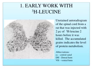
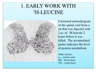
1. EARLY WORK WITH3H-LEUCINE Unstained autoradiogram of the spinal cord from a rat that was injected with 2 µc of 3H-leucine 2 hours before it was killed. The accumulated grains indicates the level of protein metabolism. Abbreviations: cc – central canal DH – Dorsal horn VH – ventral horn
2. EARLY WORK WITH 3H-LEUCINE Examples of the differences in 3H-leucine uptake by motor neurons in the cervical spinal cord in resting (left panel) and exercised (right panel) rats. RESTING EXERCISED
3. EARLY WORK WITH 3H-LEUCINE Examples of the differences in 3H-leucine uptake by neurons in the reticular formation in resting (left panel) and exercised (right panel) rats. RESTING EXERCISED
4. EARLY WORK WITH 3H-LEUCINE The results of uptake studies showed great variability between animals during and after exercise. I began to doubt the repeatability of this work, and I turned to 3H-thymidine to standardize the autoradio-graphic technique.
3H-thymidine autoradiography is the ideal technique to study cell proliferation. • The nervous system contains many proliferating glial cells in mature animals. • To our surprise, we found evidence that the small granule cells (NEURONS) in the dentate gyrus of the hippocampus are generated in the ADULT rat brain.
The Problem of Adult Neurogenesis The great Spanish neuroscientist, Santiago Ramón y Cajal, had an interest in the regeneration of the injured brain. To his disappointment, he could find no evidence of neuron multiplication in adults. That gave rise to the Dogma: NO ADULT NEUROGENESIS
THE RAT HIPPOCAMPUS This is a specialized area of the cerebral cortex in mammalian brains. Ammon’s horn (AH) contains a thin layer of pyramidal cells (pl) that form a large C-shaped curve. The dentate gyrus (DG) has a thick layer of small granule cells (gl).
Examples of granule cells (red arrows) that were generated in an adult rat brain. From: Altman, J., 1963 Autoradiographic investigation of cell proliferation in the brains of rats and cats. The Anatomical Record, 145 (4):573-591.
Adult Neurogenesis: A Systematic Research Program • To link adult hippocampal neurogenesis with embryonic, fetal, infantile, and juvenile neurogenesis, we did a systematic study of neuron development in rats. • With 3H-thymidine autoradiography Shirley Bayer established that 85% of granule cells in the dentate gyrus are generated postnatally. • Later, Bayer found that the total number of dentate granule cells increases during adulthood.
The same in a rat that was injected with 3H-thymidine on P13 and survived to P60. Note the deep placement of the labeled granule cells. The dentate granular layer in a rat that was injected with 3H-thymidine on postnatal day (P) 2 and killed on P60. Note only the most superficial granule cells are unlabeled. All the labeled cells were formed after P2. From Altman, J. 1966. Autoradiographic investigations … II. Journal of Comparative Neurology 128:431-474.
In the right dentate gyrus, the number of granule cells continually increases from around 900,000 (30 days) to over 1,275,000 (365 days). The adult-generated neurons substantially add to the population of granule cells in the hippocampus. Over 1,100 neurons are added to the population EVERY DAY between 30 and 365 days Bayer, SA et al., 1982 Science 216:890-892. Bayer, SA 1982 Experimental Brain Research 46:315-323.
While Ramón y Cajal was correct that neurons do not multiply, we showed that precursor cells of neurons, now called STEM CELLS, do multiply. • We called the layer of precursor cells in the dentate gyrus, the SUBGRANULAR ZONE.
The subgranular zone in a rat injected with 3H-thymidine on P5-P8 and killed on P9. The dentate gyrus in a rat injected with 3H-thymidine on P10 and killed 2 months later. Note the predominantly deep placement of the labeled cells.
We subsequently showed that the subgranular zone is present in the dentate gyrus of other mammals, such as the guinea pig and the cat.
GUINEA PIG SUBGRANULAR ZONE 6 DAYS 18 DAYS
CAT SUBGRANULAR ZONE 30 DAYS 60 DAYS
The complete story of hippocampal development in diagrammatic form. On Embryonic day (E)18, the neuroepithelium (NEP) is differentiated and is beginning to generate neurons that just start to migrate (arrows show trajectories). Most granule cells come from a dispersed germinal matrix, the SUBGRANULAR ZONE, not directly from the neuroepithelium. On P30, only the subgranular zone is still present AND REMAINS to generate granule cells in juvenile and adult brains.
Postnatally-forming precursors of neurons in the hippocampus are highly vulnerable and are killed with low-level X-irradiation. • This permanently interferes with adult neurogenesis.
Normal and X-irradiated Hippocampus Normal rat hippocampus at 3 months, showing the dentate gyrus (DG) with the thick granular layer (gl) and Ammon’s horn (AH) with a thin pyramidal cell layer (pl). The SELECTIVELY-reduced granular layer (arrows) in a 3 month-old rat that was given 8 doses of X-rays between P2 and P15.
We put the X-irradiated rats through a series of tests to study consequent behavioral abnormalities.
Hippocampal X-irradiation: Behavioral Studies OPEN FIELD TEST A rat is placed in the central square, and the number of squares entered during a specified time is recorded.
Hippocampal X-irradiation: Behavioral Studies The hippocampal X-irradiated animals are HYPERACTIVE in an open field.
Hippocampal X-irradiation: Behavioral Studies SPONTANEOUS ALTERNATION TEST. Will a rat re-enter an arm of a T-maze that it has already explored? Normal rats spontaneously alternate above chance level on the second trial.
Hippocampal X-irradiation: Behavioral Studies The X-irradiated animals have SHORT-TERM MEMORY DEFICITS since they readily re-enter the same arm of a T-maze in successive trials.
Hippocampal X-irradiation:Behavioral Studies The PASSIVE AVOIDANCE apparatus first shapes hungry rats to eat from a food cup when the door is opened. A day later the rats are shocked when they eat from the food cup. On the next testing day, the hungry rats are again placed in the apparatus and the door is opened. The time it takes for the rat to approach the food cup is recorded. Normal rats stay in the start box for a long time to passively avoid being shocked.
Hippocampal X-irradiation: Behavioral Studies The X-irradiated animals have LEARNING DEFICITS to avoid shock because they approach the food cup after a shorter delay than normal rats.
CONCLUSION: The “Good” News • Our research, and those of many other developmental neurobiologists, established that the regenerative capacity of the nervous system is FAR GREATER than it was believed. • Is it possible that adult-generated neurons can be coaxed into therapies to effectively remedy developmental disorders like autism, or degenerative disorders like Alzheimer’s disease?
CONCLUSION: The “Bad” News • The stem cells that give rise to neurons in adult brains are highly vulnerable and can be killed by exposure to radiation, alcohol, drugs, etc. • The absence of postnatal and adult-generated neurons affects the function of specific brain structures and leads to learning disabilities, abnormal behavior, and other disorders.
Please check our two websites for our latest research. neurondevelopment.org Here you can find pdf files of our past publications and our current work on MENTAL EVOLUTION. braindevelopmentmaps.org This is a new website that archives our histological collection of normal developing rat brains with high resolution images that can be downloaded and used for your own research.


![K 1 = [HCO 3 - ] [H + ]](https://cdn1.slideserve.com/3309135/slide1-dt.jpg)


![[D-Ala2]-Leucine enkephalin](https://cdn4.slideserve.com/8302400/d-ala2-leucine-encephalin-dt.jpg)