Download
1 / 45
450 likes | 515 Views
Explore the distinct characteristics of Ulcerative Colitis and Crohn's Disease, their clinical manifestations, diagnosis methods, and treatment options. Learn about the epidemiology, host factors, and complications associated with IBD.

E N D
Inflammatory Bowel Disease Othman alharbi, FRCPC
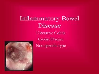
Inflammatory bowel disease (IBD) is comprised of two major disorders: • Ulcerative colitis (UC). • Crohn's disease (CD). • These disorders have both distinct and overlapping pathologic and clinical characteristics.
Epidemiology • IBD is more common in the West, but the incidence is increasing in the developing countries including Saudi Arabia. • IBD can present at any age: • The peak :15 - 30 years. • A second peak 50
High Medium Low • Inflammatory bowel disease (IBD) is comprised of two major disorders: • Ulcerative colitis (UC) • Crohn's disease (CD). • These disorders have distinct pathologic and clinical characteristics, but their pathogenesis remains poorly understood
HOST FACTORS • Genetic factors: NOD2/CARD15 • ENVIRONMENTAL FACTORS • Smoking: • Appendectomy: protect UC • Diet
Ulcerative colitis • Ulcerative colitis is characterized by recurring episodes of inflammation limited to the mucosal layer of the colon. • Start rectum then extend proximally.
Ulcerative proctitis :rectum. • Ulcerative proctosigmoiditis :rectum and sigmoid colon. • Left-sided colitis: disease that extends beyond the rectum and as far proximally as the splenic flexure. • Extensive colitis :beyond the splenic flexure. • Pancolitis: whole colon
40-50% of patients have disease limited to the rectum and rectosigmoid • 30-40% of patients have disease extending beyond the sigmoid • 20% of patients have pancolitis
The major symptoms of UC are: - diarrhea - rectal bleeding - tenesmus - passage of mucus - crampy abdominal pain
Patients with proctitis usually pass fresh blood or blood-stained mucuseither mixed with stool or streaked onto the surface of normal or hard stool • When the disease extends beyond the rectum, blood is usually mixed with stool or grossly bloody diarrhea may be noted • When the disease is severe, patients pass a liquid stool containing blood, pus, fecal matter • Other symptoms in moderate to severe disease include: anorexia, nausea, vomitting, fever, weight loss
DIAGNOSIS — • No single modalities is enough for Diagnosis. • Combination of Clinical picture, laboratory, Endoscopy, pathology.
Colonoscopy • The vascular markings are lost, petechiae, exudates, touch friability, and frank hemorrhage may be present. • Colonic involvement is continuous in ulcerative colitis, in contrast to the patchy nature of Crohn'sdisease.
Pathology: • crypt abscesses. • chronic changes including branching of crypts, atrophy of glands, and loss of mucin in goblet cells
Ulcerative colitis - complication • Hemorrhage • Perforation • Toxic megacolon (transverse colon with a diameter of more than 5,0 cm to 6,0 cm with loss of haustration) • Colon cancer
managements • Goals of therapy • Induce and maintain remission. • Ameliorate symptoms • Improve pts quality of life • Adequate nutrition • Prevent complication of both the disease and medications
managements • Role out infection • 5 ASA therapy: • Oral • Rectal • Corticosteroids: • Systemic: Prednisolone • Local acting: enema. • Immunomodulators : • Azithyoprine • Methotrexate • Anti TNF therapy
Surgery: • Severe attacks that fail to respond to medical therapy. • Complications of a severe attack (e.g., perforation, acute dilatation). • Chronic continuous disease with an impaired quality of life. • Dysplasia or carcinoma.
CD • Crohn's disease (CD) is a disorder of uncertain etiology that is characterized by transmural inflammation of the gastrointestinal tract. • CD may involve the entire gastrointestinal tract from mouth to the perianalarea. • 80% Small bowel. • 50 % ileocolitis. • 20 % colon. • 30% perianaldisease. • UGI < 5 %
CLINICAL MANIFESTATIONS • Fatigue. • Diarrhea. • Abdominal pain. • Weight loss. • Fever.
Phlegmon/abscess : • walled off inflammatory mass without bacterial infection • Fistulas : • Fistulas are tracts or communications that connect two epithelial-lined organs. • Enterovesical • Enterocutaneous • Enteroenteric • enterovaginal • Perianal disease
Severe oral involvement • aphthous ulcers. • Esophageal involvement • odynophagiaand dysphagia. • GastroduodenalCD • upper abdominal pain and symptoms of gastric outlet obstruction. • Gallstones
Extraintestinal manifestations • CD and ulcerative colitis share a number of extraintestinalmanifestations • Arthritis • Eye involvement–uveitis, iritis, and episcleritis • Skin disorders. • Others: • Primary sclerosingcholangitis, Venous and arterial thromboembolism ,Renal stones ,Bone loss and osteoporosis ,Vitamin B12 deficiency
DIAGNOSIS • The diagnosis of CD is usually established with endoscopic findings or imaging studies in a patient with a compatible clinical history.
Colonoscopy: • Endoscopic features include focal ulcerations adjacent to areas of normal appearing mucosa along with polypoid mucosal changes that give a cobblestone • Wireless capsule endoscopy
Imaging studies • small bowel follow through (SBFT) • computed tomography: CTS or CT enterography • Magnetic resonance imaging (MRI) or MRenterography
Serologic markers • Inflamatory marker : ERS, CRP • Antibody tests : • Antineutrophilcytoplasmic antibodies (pANCA) • Anti-Saccharomycescerevisiae antibodies (ASCA) • Stool markers — fecal calprotectin
Goals of therapy • Induce and maintain remission. • Ameliorate symptoms • Improve pts quality of life • Adequate nutrition • Prevent complication of both the disease and medications
Management • Role out infection • Corticosteroids: • Systemic: Prednisolone • Local acting: Budosonide. • Immunomodulators : • azithyoprine • Methotrexate
Anti TNF therapy • Surgery • Obstruction, severe perianal disease unresponsive to medical therapy, difficult fistulas, major bleeding, severe disability