Download

1 / 35
410 likes | 1.18k Views
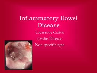
Inflammatory bowel disease . Inflammatory bowel disease. refers to two chronic diseases that cause inflammation of the intestines: ulcerative colitis and Crohn's disease . Although the diseases have some features in common, there are some important differences. Inflammatory bowel disease.

E N D
Inflammatory bowel disease • refers to two chronic diseases that cause inflammation of the intestines: ulcerative colitis and Crohn's disease. • Although the diseases have some features in common, there are some important differences.
Inflammatory bowel disease • Medical research hasn't determined yet what causes inflammatory bowel disease. But researchers believe that a number of factors may be involved, such as • environment • diet • possibly genetics
Inflammatory bowel disease • Current evidence suggests that there's likely a genetic defect that affects how our immune system works and how the inflammation is turned on and off in those people with inflammatory bowel disease, in response to an offending agent, like: • bacteria • a virus • or a protein in food
Genetics • Studies suggested that 1st degree relatives of an affected patient have a risk of IBD that is 4-20 times higher than that of general population. • The best replicated linkage region, IBD1, on chromosome 16q contains the CD susceptibility gene, NOD2/CARD15. • Having one copy of the risk alleles confers a 2–4-fold risk for developing CD, whereas double-dose carriage increases the risk 20–40-fold.
Etiology • Mutations within the NOD2/ CARD15 gene contribute to CD susceptibility. • Functional studies suggest that inappropriate responses to bacterial components may alter signaling pathways of the innate immune system, leading to • the development and persistence of intestinal inflammation. • Initiating pathogen? • Infectious? • ? Possibly non-pathogenic commensal enteric flora
Pathogenesis • The mucosa of CD patients is dominated by Th1 (T helper), which produce interferon-γ and IL-2. • In contrast, UC dominated by Th2 phenotype, which produce transforming growth factor (TGF-) and IL-5. • Activation of Th1 cells produce the down-regulatory cytokines IL-10 and TGF-.
Ulcerative colitis – microscopic features • Process is limited to the mucosa and submucosa with deeper layer unaffected • Two major histologic features: - the crypt architecture of the colon is distorted - some patients have basal plasma cells and multiple basal lymphoid aggregates
Ulcerative colitis • is an inflammatory disease of the large intestine, also called the colon. In ulcerative colitis, the inner lining - or mucosa - of the intestine becomes inflamed and develops ulcers • is often the most severe in the rectal area, which can cause frequent diarrhea.
Ulcerative colitis – macroscopic features • 40-50% of patients have disease limited to the rectum and rectosigmoid • 30-40% of patients have disease extending beyond the sigmoid • 20% of patients have a total colitis • Proximal spread occurs in continuity without areas of uninvolved mucosa
Ulcerative colitis – macroscopic features • Mucosa is : - erythematous, has a granular surface that looks like a sand paper • In more severe diseases: - hemorrhagic, edematous and ulcerated • In fulminant disease a toxic colitis or a toxic megacolon may develop ( wall become very thin and mucosa is severly ulcerated)
Ulcerative colitis – clinical presentation • The major symptoms of UC are: - diarrhea - rectal bleeding - tenesmus - passage of mucus - crampy abdominal pain
Ulcerative colitis – clinical presentation • Patients with proctitis usually pass fresh blood or blood-stained mucuseither mixed with stool or streaked onto the surface of normal or hard stool • When the disease extends beyond the rectum, blood is usually mixed with stool or grossly bloody diarrhea may be noted • When the disease is severe, patients pass a liquid stool containing blood, pus, fecal matter • Other symptoms in moderate to severe disease include: anorexia, nausea, vomitting, fever, weight loss
Ulcerative colitis - complication • Hemorrhage • Perforation • Stricture • Toxic megacolon (transverse colon with a diameter of more than 5,0 cm to 6,0 cm with loss of haustration)
Crohn’s disease • Crohn's disease differs from ulcerative colitis in the areas of the bowel it involves - it most commonly affects the last part of the small intestine and parts of the large intestine. • Crohn's disease isn't limited to these areas and can attack any part of the digestive tract • Crohn's disease generally tends to involve the entire bowel wall
Crohn’s disease – macroscopic features • Can affect any part of GI tract from the mouth to the anus • 30-40% of patients have small bowel disease alone • 40-55% of patients have both small and large intestines disease • 15-25% of patients have colitis alone • In 75% of patients with small intestinal disease the terminal ileum in involved in 90%
Distribution of gastrointestinal Crohn's disease. Based on data from American Gastroenterological Association.
Crohn’s disease – macroscopic features • CD is a transmural process • CD is segmental with skip areas in the midst of diseased intestine • In one –third of patients with CD perirectal fistulas, fissures, abscesses, anal stenosis are present
Crohn’s disease – macroscopic features • mild disease is characterized by: aphtous or small superficial ulcerations • In more active disease: stellate ulcerations fuse longitudinally and transversely to demarcate island of mucosa that are histologically normal • Cobblestone appearance is characteristic of CD (both endoscopically and by barium radiography)
Crohn’s disease – macroscopic features • Active CD is characterized by focal inflammation and formation of fistula tracts • The bowel wall thickens and becomes narrowed and fibrotic, leading to chronic, recurrent bowel obstruction
Crohn’s disease – macroscopic features • Aphtoid ulceration and focal crypt abscesses with loose aggregation of macrophages which form granulomas • Transmural inflammation that is accompanied by fissures that penetrate deeply into the bowel wall
Crohn’s disease – sign and symptoms • Ileocolitis - right lower quadrant pain and diarhhea - palpable mass, fever and leucocytosis - pain is colickly and relieved by defecation • Jejunoileitis - inflammatory disease is associated with loss of digestive and absorptive surface
Crohn’s disease – sign and symptoms Colitis and perianal disease - low grade fever, malaise, diarrhea, crampy abdominal pain, sometimes hematochezia - pain is caused by passage of fecal material through narrowed and inflamed segments of large bowel Gastroduodenal disease - nusea, vomiting, epigastric pain - second portion of duodenum is more commonly involved than the bulb
IBD is associated with variety of extraintestinal menifestation. Almost one-third of the patients have at least one.
Extraintestinal manifestation Dermatologic 1. Erythema nodosumoccurs in up to 15% of CD patients and 10% of UC patients The lesions of EN are hot, red, tender nodules measuring to 5cm in diameter and are found on the anterior surface of the legs, ankles, calves, thighs and arms 2. Pyoderma gangrenosum (PG) is seen in 1 to 12% of UC patients and is less common in CD colitis. PG may occur years before the onset of bowel symptoms. Lesions are common on the dorsal surface of the feet and legs but may occur on the arms, chest and even face.
Extraintestinal manifestation Rheumatologic Peripherial arthritis developes in 15 to 20% of IBD patients, is more common in CD. It is asymmetric, polyarticular and migratory. Most often affects large joints of the upper and lower extremities Ankylosing spondylosis (AS) occurs in 10% of IBD. Sacroilitis is symetrical, occurs equally in UC and CD, often asymptomatic
Extraintestinal manifestation Ocular The incidence of ocular complications in IBM patients is 1 to 10% The most common is conjunctivitis, anterior uveitis, episcleritis Symptoms include: ocular pain, photophobia, blurred vision, headache
Extraintestinal manifestation Urologic The most frequent genitourinary complications are: calculi, ureteral obstruction, fistulas The highest frequency of nephrolithiasis (10-20%) occurs in patients with CD.
Patients with IBD have an increased prevelance of osteoporosis secondary to vitamin D deficiency, calcium malabsorbtion, malnutrition, corticosteroid use More common cardiopulmonary manifestations include endocarditis, myocarditis, pleuropericarditis and interstitial lung disease.