SUBMANDIBULAR REGION I
280 likes | 525 Views
SUBMANDIBULAR REGION I. By Prof. Saeed Makarem. TOPICS. Suprahyoid muscles: origin, insertion, nerve supply and actions. Tongue muscles. Lingual artery : origin, course, parts and branches. SUPRAHYOID MUSCLES. 1- Digastric. 2- Stylohyoid 3- Mylohyoid. 4- Geniohyoid.

SUBMANDIBULAR REGION I
E N D
Presentation Transcript
Prof. makarem SUBMANDIBULAR REGION I By Prof. Saeed Makarem
TOPICS • Suprahyoid muscles: origin, insertion, nerve supply and actions. • Tongue muscles. • Lingual artery: origin, course, parts and branches. Prof. makarem
SUPRAHYOID MUSCLES • 1- Digastric. • 2- Stylohyoid • 3- Mylohyoid. • 4- Geniohyoid. Prof. makarem
DIGASTRIC MUSCLE • Origin & Insertion: • Posterior belly arises from the medial surface of the mastoid process, then it passes downward and forward crossing the carotid sheath, and ends in the intermediate tendon. The intermediate tendon pierces the Stylohyoid insertion and is held in position by a loop of deep fascia, which binds the tendon down to the hyoid bone. Prof. makarem
DIGASTRIC MUSCLE DIGASTRIC MUSCLE • Anterior belly runs forward and medially and is attached to the digastric fossa in the lower border of the body of the mandible, near the median plane. • Nerve supply: • posterior belly: facial nerve( 2nd) • anterior belly: nerve to the mylohyoid (branch of the mandibular division of the trigeminal nerve)-(1st pharyngeal arch) • Action: elevation of the hyoid bone Prof. makarem
STYLOHYOID MUSCLE • This is a small muscular slip that passes along the upper border of the posterior belly of digastric muscle. • Origin: from the styloid process. • Insertion: At the junction of the body & the greater cornu of hyoid. • Near its insertion, it is pierced by the intermediate tendon of the digastric muscle. • Nerve supply: Facial nerve, (2nd) • Action: elevates the hyoid bone. Prof. makarem
SUPERFICIAL RELATIONS OF THE DIGASTRIC 5 muscles, 2 glands, one vein & one bone • Platysma. • Sternomastoid. • Splenius capitis. • Longissimus capitis. • Parotid gland. • Submandibular gland. • Stylohyoid. • Retromandibular vein. • Mastoid process. Prof. makarem
DEEP RELATIONS • Deep to the anterior belly of the digatric is the mylohyoid. • Deep to the posterior belly of the digastric are: • Obliques superiorius, • occipital artery, • rectus capitis lateralis, • transverse process of atlas, • accessory nerve, • internal jugular vein, • hypoglossal nerve, • internal and external carotid arteries. • facial and lingual arteries • hyoglossus. Prof. makarem
MYLOHYOID MUSCLE Flat, triangular muscle • Origin: • Mylohyoid line of the mandible. • Insertion: • Posterior fibers into the body of the hyoid bone. • Anterior fibers into the mylohyoid raphe which extends from the symphysis menti to the body of the hyoid bone. • Nerve supply: • mylohyoid branch of the inferior alveolar nerve. Prof. makarem
Action: the two mylohyoid muscles form a muscular sheet that supports the tongue and the floor of the mouth. • When the mandible is fixed, they elevate the floor of the mouth and the hyoid bone during the first stage of swallowing. • When the hyoid bone is fixed, it assists the depression of the mandible and the opening of the mouth. Prof. makarem
GENIOHYOID MUSCLE The geniohyoid muscle is a slender muscle, superior to the mylohyoid. • Origin: from the inferior mental spine, behind the symphysis menti. • Insertion: anterior surface of the body of the hyoid bone. • Action: elevates the hyoid bone and draws it forward; or it depresses the mandible. • Nerve supply: first cervical nerve through the hypoglossal nerve. Prof. makarem
MUSCLES OF THE TONGUE • The muscles of the tongue are divided into two types: intrinsic and extrinsic. • The intrinsic musclesare confined to the tongue and are not attached to bone. • They consist of longitudinal, transverse, and vertical fibers. • Nerve supply: Hypoglossal nerve. • Action:Alter the shape of the tongue. Prof. makarem
Extrinsic Muscles of the Tongue • The extrinsic muscles are attached to bones and the soft palate. • They are: • the genioglossus, • the hyoglossus, • the styloglossus, and • the palatoglossus. Prof. makarem
Insertion: • superior fibers - to the tip of the tongue • middle fibers - to the dorsum of the tongue • inferior fibers - to the body of the hyoid bone • Nerve supply: hypoglossal nerve. GENIOGLOSSUS MUSCLE The genioglossus muscle is a fan-shaped muscle, extends backward into the tongue. • Origin: from the superior mental spine, behind the symphysis menti of the mandible. Action: draws the tongue forward and deflects the tip of the tongue to the opposite side. Simultaneous contraction of the two muscles protrude the tongue in the midline. Prof. makarem
GENIOGLOSSUS Prof. makarem
Insertion: • The fibers run downward and forward on the lateral surface of the superior constrictor muscle. • On reaching the interval between the superior and the middle constrictor muscles, the styloglossus passes forward to enter the sides of the tongue. STYLOGLOSSUS MUSCLE The styloglossus muscle is a long, slender muscle. • Origin:from the styloid process. • Action: draws the tongue upward and backward. • Nerve supply: hypoglossal nerve. Prof. makarem
HYOGLOSSUS MUSCLE The hyoglossus is a flat, quadrilateral muscle, superficial to the mylohyoid. • Origin: from upper border of the body and greater cornu of the hyoid bone. • Insertion: to the side of the tongue. • Action: depression of tongue. • Nerve supply: hypoglossal nerve. Prof. makarem
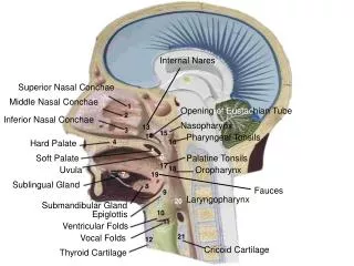
STRUCTURES ON THE LATERAL SURFACE OF THE HYOGLOSSUS MUSCLE • From above downwards: • Lingual nerve. • Submandibular ganglion. • Deep part of the submandibular salivary gland. • Submandibular duct. • Hypoglossal nerve. • Suprahyoid artery. Prof. makarem
Branches • Dorsal lingual branches are two or three in number and ascend to the dorsum of the tongue. • The sublingual artery supplies the sublingual salivary gland and neighboring structures. • Suprahyoid artery :from the beginning of the artery and runs superficial to Hyoglossus • Deep lingual artery: runs on the under surface of the tongue under its mucous membrane • It runs forward, forming an upward loop, which is crossed by the hypoglossal nerve. • It then proceeds forward deep to the hyoglossus muscle to supply the tip of the tongue. LINGUAL ARTERY The lingual artery arises from the external carotid artery opposite the tip of the greater cornu of the hyoid bone. Prof. makarem