Download
1 / 53
530 likes | 663 Views
THE DOCTORS GUIDE TO PATIENT SURVIVAL AFTER ACUTE AORTIC DISSECTION. Mark J. Russo, MD, MS Co-Director, Center for Aortic Diseases University of Chicago. THE NUMBERS. Incidence - uncertain 5,000 – 15,000 cases/year in U.S. Likely higher (not reportable condition, few autopsies now)

E N D
THE DOCTORS GUIDE TO PATIENT SURVIVAL AFTER ACUTE AORTIC DISSECTION Mark J. Russo, MD, MS Co-Director, Center for Aortic Diseases University of Chicago
THE NUMBERS • Incidence - uncertain • 5,000 – 15,000 cases/year in U.S. • Likely higher (not reportable condition, few autopsies now) • Autopsy series – 0.2% autopsies • Males 2-5x > females • Ascending dissections: 50-55 years old • <40 years: Marfan, pregnancy, AV disease • Descending dissections: 60-70 years old
NATURAL HISTORY • 1934 Shennan: >300 cases autopsies reviewd 40% acute ascending dissections died suddenly None lived > 5 weeks • 1972 Anagnostopoulos : 973 pts w untreated proximal and distal dissections 50% died with 48 hours 84% died within 1 month
NATURAL HISTORY • As many as 40% die before reaching the hospital • Mortality increases 1-3% per hour • At 48 hours, 50% are dead • At 2 weeks 75-90% are dead
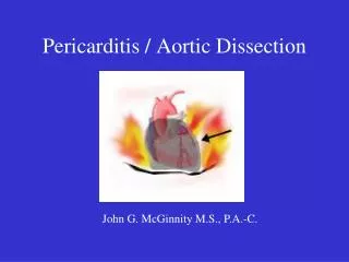
MECHANISM • Initiating event may be is a primary rupture of the intima with secondary dissection of the media -- OR -- • Hemorrhage within the media and subsequent rupture of the overlying intima.
MECHANISM • Blood flow is redirected from the “true lumen” of the aorta into a “false lumen”
MECHANISM • As a result, dissection propagates in “dissection plane” separating the intima from the overlying adventitia • Usually the dissection proceeds distally/ retrograde/direction of blood flow
MECHANISM • Dissection may shear off or extend into branch arteries -> complications
COMPLICATIONS - MALPERFUSION Stroke (3-13%) arm ischemia (25-60%) arm ischemia (25-60%) Paralysis (2-9%) kidney dysfunction (25%-75%) bowel ischemia (10%-20%) MI (5-10%) Tamponade (10%) leg ischemia (25-60%)
CLASSIFACTION Debakey I II IIIaIIIb Stanford A A B B
YOUR ROLE • Diagnosis • Suspicion • Treatment • Medical Management - Always • Consult a Surgeon - Always
TIME MATTERS • As many as 40% die before reaching the hospital • Mortality increases 1-3% per hour • At 48 hours, 50% are dead • At 2 weeks 75-90% are dead
Most important factor leading to a correct diagnosis is a high clinical suspicion
PRESENTATION • Pain - severe chest, back, and/or limb – 90% • Severe uncontrolled hypertension – 50-60% • Loss of consciousness (syncope) – 15% • Weakness • Difficulty walking • Slurred speech • Blurry/loss or vision
PAIN CHARACTERISTICS • Occurs in 90% of cases • Ripping, tearing • Migratory • Never experienced before • Restless, sense of doom • Most Severe at Onset • Anterior Pain: Proximal Dissection • Posterior Pain: Distal Dissection • Migratory Pain
PAIN CHARACTERISTICS • Chest pain – 2/3 • Back pain – 1/2 • Abdominal pain – 1/3 • Painless dissection is relatively uncommon (6.4%) • Presenting symptoms of syncope, heart failure, or stroke were seen more often in this group. • Pain in these locations usually due to other more common disorders (MI, pneumonia, pleurisy, pulmonary embolism, pneumothorax, ulcer, cholecystitis, pancreatitis) BUT….
Must consider aortic dissection in cases without other confirmed cause of pain
RISK FACTORS • Hypertension - Present in 70-90% of dissections, but 20-40% of adults • Aortic aneurysm – 13% • Family history of aortic disease – 19% • Connective tissue diseases - Marfans (2%), Ehlers-Danlos, Lowy-Dietz • Bicuspid aortic valve – 1% • Aortic coarctation • Turner syndrome • Cardiac intervention – CABG, AVR, Cath (2%) • Pregnancy • Trauma • High Intensity weightlifting • Crack – 37% in an inner city population, usually < 12 hours after last use
PHYSICAL EXAMINATION • Acutely ill • Tachycardia • Hypertension – particularly if severe HTN • Results catecholamines, renal ischemia • Hypotension (20%) • Due to acute complications • Widen Pulse Pressure • Aortic insufficiency: (50-60% ascending dissections) • Differential pressure from Left to Right Arm (when dissection is distal to BCA) • Pulse deficits: (60% ascending dissections) • May change over time
D-DIMER • D-Dimer is an important and well-known marker for pulmonary embolism (PE), especially in outpatients and the emergency department. • Also a biomarker for aortic dissection, because of the associated large intramural hematoma often present in aortic dissections. • Initial D-Dimer value in symptomatic patients with concerns for aortic dissection: • D-Dimer < 0.5 μg/ml: Thoracic Ascending Aortic Dissection unlikely • D-Dimer >1.6 μg/ml: Thoracic Ascending Aortic more likely, • proceed with aortic imaging with CT C/A/P with IV contrast or TEE • Thoracic Ascending Aortic Dissection (TAAD) elevates D-Dimer Earlier Than Pulmonary Embolus
CXR • Mediastinal widening - 63% w type A dissections • Pleural effusion - 19% of dissections • Other findings: • widening of the aortic contour, • displaced calcification, • aortic kinking, and • opacificationof the aorticopulmonarywindow • Normal - 11%
CXR Features of acute type A dissection,
CXR • Features of acute type A dissection, • Widened mediastinum
CXR • Features of acute type A dissection, • Widened mediastinum • Rightward tracheal displacement
CXR • Features of acute type A dissection, • Widened mediastinum • Rightward tracheal displacement • Irregular aortic contour with loss of the aortic knob
CXR • Features of acute type A dissection, • Widened mediastinum • Rightward tracheal displacement • Irregular aortic contour with loss of the aortic knob • Indistinct aortopulmonary window • Left pleural effusion
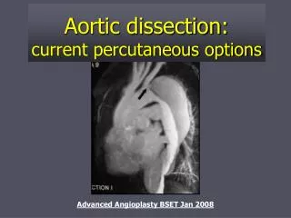
IMAGING - PURPOSE • Dissection flap • Dilated aorta • Aortic insufficiency • Pericardial effusion • Involvement of the ascending aorta • Branch vessel or coronary artery involvement • Extent of dissection and the sites of entry and reentry • Thrombus in the false lumen
IMAGING - OPTIONS • Most have multiple imaging studies performed • mean of 1.83 per patient • Transthoracic echocardiogram – 33% • Transesophageal echocardiogram - 33% • Computed tomography- 61% • Aortography – 4% • Magnetic resonance imaging – 2%
CT SCAN • Sensitivity - 83 and 98%; specificity - 87 and 100% • Advantages • Availability at most hospitals • Identification of intraluminal thrombus and pericardial effusion • Disadvantages • Intimal flap is seen in < 75% • Site of entry is rarely identified • Nephrotoxic iodinated contrast is required • No capability to assess for aortic insufficiency
CT False Lumen [tear] True Lumen
Transthoracic Echo • Sensitivity and specificity inferior to CT, MRI, and TEE • Advantages • Noninvasive • Fast, low risk • Intimal flap may be seen in the proximal aorta in some patients • Useful for the assessment of cardiac complications of dissection, including aortic insufficiency, pericardial effusion/tamponade, and RV function. • Disadvantages - inability to adequately visualize the distal ascending, transverse, and descending aorta in a substantial majority of patients
Transesophageal Echo • Sensitivity 97 to 99 percent; , the specificity 77 to 85 percent • Advantages • Rapid; useful in patients too unstable for CT/MRI • True and false lumens can be identified • Intimal dissection flaps can be identified • Thrombosis in the false lumen, pericardial effusion, concomitant aortic regurgitation, and the proximal coronary arteries can be readily visualized. • Disadvantages • Requires esophageal intubation • Requires the availability of experienced operators (both physicians and technicians) • Inability to visualize the upper portion of the ascending aorta due to the interposed trachea (between the aorta and esophagus).
MRI • Sensitivity and specificity of MRI were both 98% • Advantages • 85% sensitivity for identification of the site of entry • MR contrast agents have a more favorable safety profile than iodinated contrast agents. • ability to assess branch vessels. • Disadvantages • Long study • limited applicability (MRI cannot be performed in patients with claustrophobia, pacemakers, or certain types of aneurysm clips or metallic ocular/auricular implants). • not readily available on an emergency basis at many institutions • concerns about patient monitoring and relative patient inaccessibility during prolonged scanning • Unable to assess for aortic insufficiency
MANAGEMENT • Mean arterial pressure of 60-75 mmHg: • 1st line treatment: Beta blockers (egesmolol, propranolol, or labetalol) • If there is a contraindication to beta blockers, calcium-channel blockers (eg verapamil and diltiazem) can be used • For refractory hypertension: Nitroprusside, in addition to a beta- or calcium-channel blockers. • DO NOT USE: Hydralazine or minoxidil or beta-blockers with intrinsic sympathomimetic action (eg, acebutolol, pindolol)
MANAGEMENT • It is not your job to make a definitive diagnosis • If you suspect….call a surgeon • Call a surgeon • Call a surgeon
RITTERS RULES • Life-saving reminders to recognize, treat and prevent thoracic aortic dissection • Named for actor John Ritter, who died of a thoracic aortic dissection, Ritter Rules combine knowledge with action. • Address urgency, symptoms, who is most at risk and which imaging tests
URGENCY • Thoracic aortic dissection is a medical emergency. • The death rate increases 1% every hour the diagnosis and surgical repair are delayed.
PAIN • Severe pain is the #1 symptom. • Sudden onset of severe pain in the chest, stomach, back or neck. • is likely to be sharp, tearing, ripping, moving or so unlike any pain you have ever had that you feel something is very wrong.
MISDIAGNOSIS • Aortic dissection can mimic heart attack. • If a heart attack or other important diagnosis is not clearly and quickly established, then aortic dissection should be quickly considered and ruled out, • particularly if a patient has a family history or features of a genetic syndrome that predisposes the patient to an aortic aneurysm or dissection.
IMAGING • Get the right scan to rule out aortic dissection. • Only three types of imaging studies can identify aortic aneurysms and dissections: CT, MRI and transesophageal echocardiogram. • A chest X-ray or EKG cannot rule out aortic dissection.