Download
1 / 60
630 likes | 830 Views
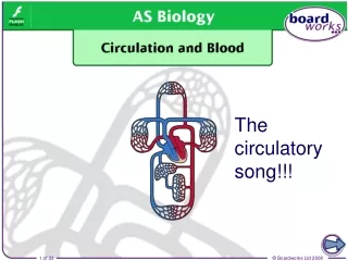
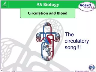
The circulatory song!!!. 3:05. The giraffe and the problem with its long neck!. Why need a transport system?. Single-celled organisms, such as bacteria and amoeba ( below ), can obtain nutrients and excrete waste simply by diffusion. nutrients. waste products.

E N D
3:05 The giraffe and the problem with its long neck!
Why need a transport system? Single-celled organisms, such as bacteria and amoeba (below), can obtain nutrients and excrete waste simply by diffusion. nutrients waste products Multi-cellular organisms, such as insects, fish and mammals, require a more specialized transport system. Why is this?
Surface area to volume ratio In larger organisms, diffusion of substances would occur far too slowly to enable them to survive: the rate of diffusion increases with the square of the distance it has to travel. This is not just because of its size, however: more important is an organism’s surface area to volume ratio. Single-celled organisms have a very large surface area to volume ratio, because the diffusion path is so short.
Components of circulatory systems Multi-cellular animals overcome the limitations of diffusion by having a specialized circulatorysystem. This comprises: • a heart • a fluid in which substances are transported • vessels through which the fluid can flow. The two types of circulatory system are open (e.g. molluscs, arthropods) and closed (e.g. vertebrates, a few invertebrates).
Top trump cards • Use your background reading sheets and text book to fill out the information cards. These are outlines to top trump cards. • Decide on which answer is going to be better than the other and why. • If you are not sure what the terms mean- look them up!!!
Open circulatory systems An open circulatory system consists of a heart that pumps a fluid called haemolymph through short vessels and into a large cavity called the haemocoel. In the haemocoel, the haemolymph directly bathes organs and tissues, enabling the diffusion of substances. heart haemocoel When the heart relaxes, the haemolymph blood is sucked back in via pores called ostia. Haemolymph moves around the haemocoel due to the movement of the organism.
Closed circulatory systems In a closed circulatory system, blood is fully enclosed within blood vessels at all times. From the heart, blood is pumped through a series of progressively smaller vessels. In the smallest vessels, capillaries, substances diffuse in and out of the blood and into cells. heart capillaries Blood then returns to the heart via a series of progressively larger vessels.
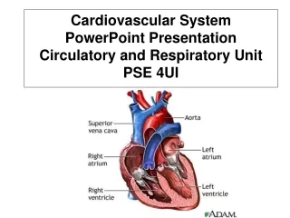
The human heart The heart is a muscular organ located between the lungs in the centre of the chest (thorax), and is about the size of a fist. It pumps blood continuously around the body. An organism can lose consciousness within just a few seconds if the brain is deprived of blood. In foetuses, the heart begins beating about 5–6 weeks after conception.
Cardiac muscle The heart mainly consists of cardiac muscle tissue, which like smooth muscle (but not skeletal muscle), contracts involuntarily. Cardiac muscle is made up of cells that are connected by cytoplasmic bridges. This enables electrical impulses to pass through the tissue. It contains large numbers of mitochondria and myoglobin molecules.
Cardiac output The amount of blood pumped around the body is called the cardiac output, and depends on two factors: • the stroke volume – the volume of blood pumped by the left ventricle in each heart beat. A typical value for an adult at rest is 75ml. • the heart rate – the number of times the heart beats per minute. A typical value for an adult at rest is 70bpm. cardiac output = stroke volume × heart rate A typical resting cardiac output is 4–6 litres per minute. This can rise to as much as 40 litres per minute in highly trained endurance athletes.
Pacemaker cells of the heart The heart can beat without any input from the nervous system as longs as its cells stay alive. This is due to myogenic contraction. Muscle cells (myocytes) in the heart have a slight electrical charge across their membrane. They are polarized. When the charge is reversed, they are said to be depolarized and this causes them to contract. Depolarization is initiated in a region of the heart called the sinoatrial node (SAN) – also known as the pacemaker – which is in the wall of the right atrium.
Artificial pacemakers Artificial pacemakers are devices implanted in people whose heart’s electrical conduction system is not working properly. Problems include the SAN not firing, and the blockage or disruption of impulses between the SAN and AVN, or in the bundle of His. Pacemakers monitor the heart’s electrical activity and stimulate the ventricles or atria to contract when necessary. Impulses are transmitted down electrodes implanted in the muscular walls.
What are electrocardiograms? The electrical activity of the heart can be monitored by an electrocardiograph. Several electrodes are attached to specific places on a person’s chest and limbs. These detect changes in polarization in the heart by measuring current at the skin surface. The leads are connected to a machine that draws an electrocardiogram (ECG).
ECG in diagnosis ECGs are used to diagnose problems with the heart, as variations in different components of the trace can indicate a disease or other abnormality. An ECG may be taken while the patient is relaxed or it may be taken before, during and after exercise. This is called a ‘stress test’ and usually involves the patient exercising on a treadmill while attached to an ECG machine.
Varicose veins If a vein wall becomes weakened, valves may no longer close properly. This allows backflow of blood, causing the vein to become enlarged and bumpy, and become varicose. This usually happens in superficial veins, near the skin surface in the lower legs, as opposed to deep veins, which lie underneath muscles. Varicose veins can be surgically removed without affecting blood flow, as most blood is returned to the heart by deep veins.
Maintaining high blood pressure Blood pressure is the main force that drives blood from the heart around the body. • During systole (heart contraction), blood is pumped through the aorta and other arteries at high pressure. The elastic fibres of arteries enable them to expand and allow blood through. • During diastole (heart relaxation), the blood pressure in the arteries drops. The elastic recoil of the artery walls help force the blood on. As blood moves through smaller arterioles into capillaries, and then into venules and veins, its velocity and pressure drop continuously.
What is blood? Blood is a specialized transport medium that is also considered a special type of connective tissue. An average adult has 4–6 litres of blood. Blood has a range of functions such as: • transport • defence • thermoregulation • maintaining pH of body fluids.
Features of erythrocytes What are the specialized features of an erythrocyte? flattened, biconcave discshape: ensures large surface area to volume ratio for efficient gas exchange large amount of haemoglobin:for transporting oxygen no nucleus or organelles: maximises space for haemoglobin, so more oxygen can be transported diameter (6–8µm) largerthan capillary diameter: slows blood flow to enable diffusion of oxygen