Download
1 / 58
1.59k likes | 5.57k Views
Blood-Hematopoiesis-Lymphatics 2013. The Megaloblastic Anemias: Vitamin B 12 & Folate Deficiency. William Kern, MD Director, Clinical Hematopathology william-kern@ouhsc.edu.

E N D
Blood-Hematopoiesis-Lymphatics 2013 The Megaloblastic Anemias:Vitamin B12 & Folate Deficiency William Kern, MD Director, Clinical Hematopathology william-kern@ouhsc.edu
Downloading or copying any of the photographs, images or diagrams from this presentation for any purpose other than studying for BHL is prohibited
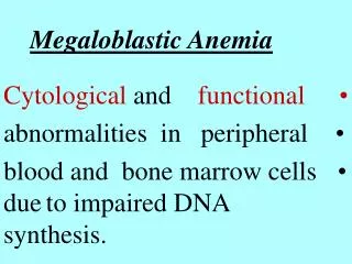
Megaloblastic Anemias: Pathophysiology • Impaired DNA synthesis: • Delayed nuclear maturation • DNA abnormalities, including chromatin strand breaks • Ineffective hematopoiesis: • Hyperproliferative marrow, but many cells die within marrow Note: All dividing cells in body affected-not just bone marrow
Megaloblastic Anemia • All dividing cells in body affected: • Bone marrow: Anemia or pancytopenia • Gastrointestinal tract: Mucosal atrophy, glossitis • Skin: Thin skin, fine hair • Others
Megaloblastic Anemias: Causes • Cobalamin (Vitamin B12) deficiency • Folic acid (folate) deficiency • Anti-folate drugs: Methotrexate, trimethoprim/sulfa antibiotics • Other drugs: Nucleoside inhibitors (zidovudine [AZT])
Terminology • Macrocytic anemia: Anemia with increased MCV: • May or may not be megaloblastic • Megaloblastic anemia: Anemia due to impaired DNA synthesis: • Usually but not always macrocytic • Pernicious anemia: Cobalamin (B12) deficiency due to autoimmune gastritis: • Subset of megaloblastic anemia
Folate deficiency Cobalamin deficiency Anti-folate drugs Liver disease Reticulocytosis Myelodysplasia Alcoholism Hypothyroidism Megaloblastic vs. Macrocytic Anemias Megaloblastic: Macrocytic: Distinction between macrocytic anemia and megaloblastic anemia is critical!
Folic Acid & Cobalamin • Folic acid = pteroylglutamic acid: • Important in transfer of one carbon fragments in many chemical reactions • Cobalamins: Several compounds • Vitamin B12 = cyanocobalamin; not naturally occurring • “Cobalamin” and “vitamin B12” used synonymously
Folic Acid • Dietary sources: Vegetables, fruit, cereals, dairy products • Heat labile; destroyed by cooking: • Fresh fruits & vegetables = main source • Daily requirement ~50 micrograms; body stores ~5-10 milligrams: • Stores can be depleted in a few months • Enterohepatic circulation Grain products now routinely supplemented with folic acid
Causes of Folate Deficiency • Inadequate diet: Most common • No fresh fruits and vegetables • Alcoholism • Malabsorption: Less common • Celiac sprue, tropical sprue, extensive bowel resection, regional enteritis • Uncommon causes: • Hemodialysis, anti-epileptic drugs, oral contraceptives (rare), nitrous oxide
Folic Acid: Increased Need • Chronic hemolytic anemias: • Hemoglobinopathies: Sickle cell diseases, others • Hereditary spherocytosis • Psoriasis • Pregnancy
Folic Acid & Pregnancy • Folate deficiency in pregnant women associated with increased risk of neural tube defects in infants • Adequate folate intake important for all women who are pregnant or trying to become pregnant In 1998, FDA mandated that all grain products in U.S. be supplemented with folate
Cobalamin • Primary sources: Meat, eggs, dairy products • Daily requirement: ~0.1 microgram/day • American diet: ~5-15 micrograms/day • Body stores ~2-4 milligrams: • Body stores adequate for years
Cobalamin Deficiency: Causes • Malabsorption: Most common cause • Dietary deficiency: Extremely rare in U.S.: • Strict vegans • Infants of vegetarian mothers • Congenital: Rare • Deficiency of intrinsic factor or transcobalamin II
Cobalamin Absorption • Complex; several steps where process can break down • Key step:Intrinsic factor (IF) produced by gastric parietal cells binds to B12 • IF-B12 complex efficiently absorbed by specific receptors in terminal ileum: • <2% of B12not bound to IF absorbed
Cobalamin Absorption (1) • B12 in food bound to protein • Gastric acid & pepsin digest B12 off of food protein • Haptocorrin (R-binders) from salivary glands binds to B12, blocking IF Robbins Pathologic Basis of Disease
Cobalamin Absorption (2) • Pancreatic enzymes digest R-binders off of B12 • IF now binds to B12 • IF-B12 complex absorbed in terminal ileum • B12 transported in blood by transcobalamin II Robbins Pathologic Basis of Disease
Cobalamin Malabsorption • Food-cobalamin malabsorption: Most common cause, but most cases subclinical • Pernicious anemia: Most common cause of overt cobalamin deficiency • Resection or disease of terminal ileum • Inflammatory bowel disease • Pancreatic insufficiency • Blind loop syndrome • Fish tapeworm (Diphyllobothrium latum)
Food-Cobalamin Malabsorption • Inability to absorb B12 bound to food protein: • Able to absorb crystalline (free) B12 • Deficiency of pepsin & gastric acid • Overall most common cause of B12 deficiency • Majority of cases subclinical rather than overt • Clinical significance debated
Food-Cobalamin Malabsorption: Causes • Gastric surgery (including bariatric surgery) • Chronic gastritis: Due to Helicobacter pylori and others • Antacids (H2-blockers, proton pump inhibitors) may exacerbate • Metformin also exacerbates
Pernicious Anemia • Autoimmune chronic gastritis with destruction of gastric parietal cells • Loss of intrinsic factor production: inability to absorb B12 • Occurs in all ethnic groups: • Highest incidence in Scandinavian, English, Scottish, Irish • Usually older population • African-Americans: Common in young women
Pernicious Anemia: Gastric Biopsy Normal Stomach Pernicious Anemia
Pernicious Anemia (PA) • Familial predisposition • Associated with other autoimmune diseases: • Thyroiditis (Hashimoto’s, Graves’ disease), Addison’s disease, vitiligo • Autoantibodies: • Anti-parietal cell antibodies: ~90% of patients, but not specific for PA • Anti-Intrinsic factor antibodies: ~50-60% of patients; specific for PA
Pernicious Anemia: Association with Gastric Tumors • Pernicious anemia associated with increased risk of gastric tumors • Carcinoid tumors appear to be highest risk • Gastric lymphomas, adenocarcinomas and other tumors also increased
Key Reactions (1) • Conversion of dUMP to dTMP by thymidylate synthetase • 5,10-methylene tetrahydrofolate required • dTMP required for DNA synthesis • Cobalamin required for regeneration of methylene-FH4 Thymidylate Synthetase dUMP dTMP dTTP DNA Methylene-FH4 FH2 Cobalamin (several intermediate steps)
Key Reactions (2) • Conversion of homocysteine to methionine by methionine synthetase • Requires methyl-FH4 and cobalamin • Allows regeneration of methylene-FH4 for DNA synthesis Methylene-FH4DNA Methyl-FH4 FH4 Homocysteine Methionine Methyl-B12 S-adenosyl-methionine (SAM) S-adenosyl-methionine (SAM) required for myelin synthesis and maintenance
Summary: Folate & Cobalamin Interaction Thymidylate Synthetase dUMP dTMP DNA Methylene-FH4 FH2 Several intermediate steps FH4 Methyl-FH4 Methionine Homocysteine Methyl-B12 Methionine Synthetase
Weakness, fatigue Painful tongue & mouth Weight loss Loss of appetite, nausea, vomiting Loose stools Pallor, “lemon yellow” color Angular cheilitis Dry, smooth skin Smooth, “beefy” red tongue Silvery or premature graying of hair Megaloblastic Anemia Symptoms: Signs:
Megaloblastic Anemia “Lemon yellow” skin “Beefy” red tongue
Megaloblastic Anemia Angular Cheilitis
Megaloblastic Anemia:Hematologic Effects • Pancytopenia common • Anemia tends to be most striking: • Hemoglobin can be ≤5 g/dL • Macrocytosis with oval macrocytes • Hypersegmented neutrophils: “Rule of Fives”: • >5 nuclear lobes, or: • ≥5% of neutrophils have 5 lobes
Megaloblastic Anemia: Blood Smear Hypersegmented Neutrophil; Macroovalocytes (↑)
Megaloblastic Anemia:Bone Marrow • Hypercellular • Marked erythroid hyperplasia • “Megaloblastic maturation”: Nucleus to cytoplasm asynchrony: • Large, immature nuclei in late erythroid precursors • Giant bands & metamyelocytes
Megaloblastic Anemia:Other Laboratory Findings • Marked increase in lactic dehydrogenase (LDH) • Increased bilirubin-predominantly unconjugated: • LDH and bilirubin increased due to intramedullary hemolysis • Haptoglobin may be decreased
Cobalamin Deficiency:Neurologic Manifestations • Highly variable; may be absent • Early: Paresthesias of distal extremities; reduced vibration & position sensation • Later: Clumsiness, ataxia, weakness, hyperreflexia, positive Babinski & Romberg signs Note: Folate deficiency is usually not associated with neurologic manifestations
Cobalamin Deficiency:Neurologic Manifestations • Pathophysiology: Myelin degeneration & loss of axons in dorsal & lateral columns of spinal cord and cerebral cortex • Both motor & sensory systems affected: • “Subacute combined degeneration” • “Combined systems disease”
Cobalamin Deficiency:Cortical Manifestations • May have predominantly cortical manifestations: • Dementia • Altered mental status • Bizarre behavior • Mental status or behavioral changes may occur in absence of neuropathy or spinal cord manifestations
Cobalamin Deficiency:Neurologic vs Hematologic Changes • Poor correlation between neurologic and hematologic changes: • Neurologic manifestations may be present without macrocytosis or anemia • Patients with severe hematologic changes may have no neurologic signs The absence of anemia or macrocytosis does not exclude neurologic disease due to cobalamin deficiency!
Cobalamin Deficiency:Neurologic Manifestations • Early: Treatment with B12 may reverse all neurologic manifestations • Long-standing changes (≥6 months) may be irreversible: • Complete response may take 6-12 months • Inappropriate treatment with folate may cause hematologic response, but may accelerate neurologic progression
Folate vs. Cobalamin Deficiency • Hematologic manifestations identical • Neurologic disease is strong indicator of cobalamin, rather than folate deficiency: • However, patients with folate deficiency may have neurologic disease due to other causes • Patients with cobalamin deficiency may have no neurologic manifestations
Serum cobalamin level Serum folate level Erythrocyte folate level Serum methylmalonic acid (MMA) Serum homocysteine level Laboratory Diagnosis of Megaloblastic Anemia Primary Lab Tests: Secondary Lab Tests:* * Helpful in borderline or equivocal cases
Laboratory Diagnosis of Megaloblastic Anemia * MMA = Methylmalonic acid
Laboratory Diagnosis of Folate Deficiency • Serum folate level labile; reflects last few meals • Erythrocyte folate more stable; reflects last 1-2 weeks: • Better index of body folate stores • Serum folate may appear falsely normal if patient fed before level drawn
Laboratory Diagnosis of Cobalamin Deficiency • Serum cobalamin is not the most sensitive test for cobalamin deficiency • Serum methylmalonic acid (MMA) more sensitive: • Increases in cobalamin deficiency • If B12 level is borderline or low normal, get MMA level
Schilling Test • Used to identify cause of B12 deficiency • Classic: Two stage • Stage I: Oral radiolabeled B12 plus IM “cold” B12 (flushing dose): • Collect urine for 24-72 hours • If abnormal, proceed to stage II • Stage II: Oral radiolabeled B12 complexed with IF; IM “cold” B12: • Repeat urine collection No longer available
Schilling Test: Interpretation • Normal: >6% of radiolabeled B12 excreted into urine • Pernicious anemia: • Stage I (No IF): <6% excreted into urine • Stage II (with IF): Corrects deficiency • Other causes of B12 malabsorption: IF fails to correct deficiency
Schilling Test: Pitfalls • Improper 24 hour urine collection: • Too little or too much collected • Failure to get flushing dose of “cold” B12 into muscle • Folate or B12 deficiency may cause malabsorption due to mucosal atrophy: • May need to repeat test after few weeks of treatment