Download
1 / 79
790 likes | 970 Views
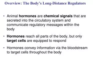
Overview: The Body’s Long-Distance Regulators. Animal hormones are chemical signals that are secreted into the circulatory system and communicate regulatory messages within the body Hormones reach all parts of the body, but only target cells are equipped to respond

E N D
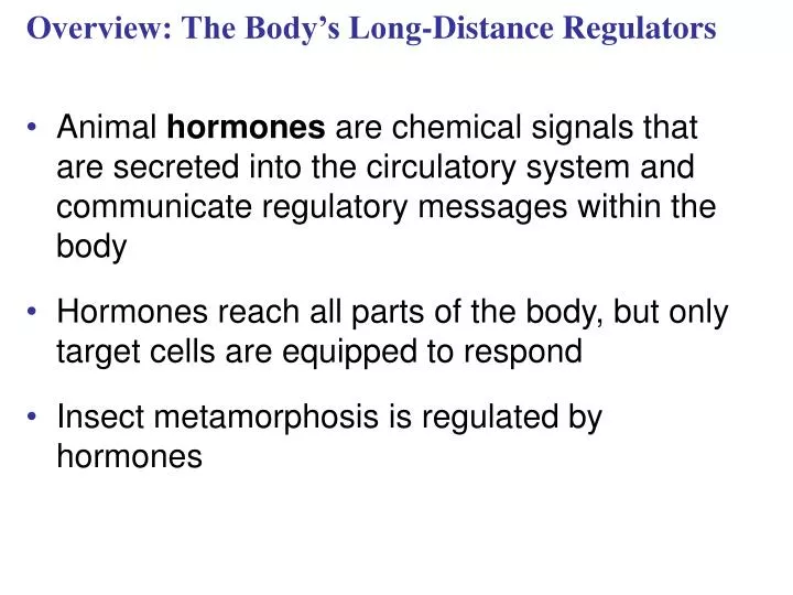
Overview: The Body’s Long-Distance Regulators • Animal hormones are chemical signals that are secreted into the circulatory system and communicate regulatory messages within the body • Hormones reach all parts of the body, but only target cells are equipped to respond • Insect metamorphosis is regulated by hormones
Chapter 45 Hormones and theEndocrine System
Types of Secreted Signaling Molecules • Secreted chemical signals include • Hormones :Endocrine signals Local regulators • Neurotransmitters • Neurohormones • Pheromones Exocrine glands
Fig. 45-2 Blood vessel Response (a) Endocrine signaling Response (b) Paracrine signaling Response (c) Autocrine signaling Synapse Neuron Response (d) Synaptic signaling Neurosecretory cell Blood vessel Response (e) Neuroendocrine signaling
Local Regulators • Local regulators are chemical signals that travel over short distances by diffusion • Local regulators help regulate blood pressure, nervous system function, and reproduction • Local regulators are divided into two types • Paracrine signals act on cells near the secreting cell • Autocrine signals act on the secreting cell itself
Fig. 45-2a Blood vessel Response (a) Endocrine signaling Response (b)Paracrine signaling Response (c) Autocrine signaling
Fig. 45-2b Synapse Neuron Response (d) Synaptic signaling Neurosecretory cell Blood vessel Response (e) Neuroendocrine signaling
Chemical Classes of Hormones • Three major classes of molecules function as hormones in vertebrates: • Polypeptides (proteins and peptides) • Amines derived from amino acids • Steroid hormones
Fig. 45-3 Water-soluble Lipid-soluble 0.8 nm Polypeptide: Insulin Steroid: Cortisol Amine: Epinephrine Amine: Thyroxine
Hormone Receptor Location: Scientific Inquiry • In the 1960s, researchers studied the accumulation of radioactive steroid hormones in rat tissue • These hormones accumulated only in target cells that were responsive to the hormones • These experiments led to the hypothesis that receptors for the steroid hormones are located inside the target cells • Further studies have confirmed that receptors for lipid-soluble hormones such as steroids are located inside cells
Researchers hypothesized that receptors for water-soluble hormones would be located on the cell surface • They injected a water-soluble hormone into the tissues of frogs
Fig. 45-4 1.The hormone triggered a response only when it was allowed to bind to cell surface receptors 2. This confirmed that water-soluble receptors were on the cell surface RESULTS MSH injected into melanocyte Melanocyte with melanosomes (black dots) Melanosomes do not disperse Melanosomes disperse Nucleus MSH injected into interstitial fluid (blue)
Cellular Response Pathways • Water and lipid soluble hormones differ in their paths through a body • Water-soluble hormones are secreted by exocytosis, travel freely in the bloodstream, and bind to cell-surface receptors • Lipid-soluble hormones diffuse across cell membranes, travel in the bloodstream bound to transport proteins, and diffuse through the membrane of target cells
Fig. 45-5-2 Fat-soluble hormone Water- soluble hormone Transport protein Signal receptor TARGET CELL OR Signal receptor Cytoplasmic response Gene regulation Cytoplasmic response Gene regulation NUCLEUS (a) (b)
Pathway for Water-Soluble Hormones Fig. 45-6-1 Epinephrine Adenylyl cyclase G protein GTP G protein-coupled receptor ATP Second messenger cAMP
Fig. 45-6-2 Epinephrine Adenylyl cyclase G protein GTP G protein-coupled receptor ATP Second messenger cAMP Protein kinase A Inhibition of glycogen synthesis Promotion of glycogen breakdown
Fig. 45-7-1 Hormone (estradiol) Pathway for Lipid-Soluble Hormones Estradiol (estrogen) receptor Plasma membrane Hormone-receptor complex
Fig. 45-7-2 Hormone (estradiol) Estradiol (estrogen) receptor Plasma membrane Hormone-receptor complex DNA Vitellogenin mRNA for vitellogenin
Multiple Effects of Hormones • The same hormone may have different effects on target cells that have • Different receptors for the hormone • Different signal transduction pathways • Different proteins for carrying out the response • A hormone can also have different effects in different species
Fig. 45-8-1 Same receptors but different intracellular proteins (not shown) Epinephrine Epinephrine receptor receptor Glycogen deposits Vessel dilates. Glycogen breaks down and glucose is released. (a) Liver cell (b) Skeletal muscle blood vessel
Fig. 45-8-2 Same receptors but different intracellular proteins (not shown) Different receptors Epinephrine Epinephrine Epinephrine Epinephrine receptor receptor receptor receptor Glycogen deposits Vessel dilates. Vessel constricts. Glycogen breaks down and glucose is released. (a) Liver cell (c) Intestinal blood vessel (b) Skeletal muscle blood vessel
Fig. 45-9 (a) (b)
Signaling by Local Regulators • In paracrine signaling, nonhormonal chemical signals called local regulators elicit responses in nearby target cells • Types of local regulators: • Cytokines and growth factors • Nitric oxide (NO) • Prostaglandins: • Prostaglandins help regulate aggregation of platelets, an early step in formation of blood clots
Concept 45.2: Negative feedback and antagonistic hormone pairs are common features of the endocrine system • Hormones are assembled into regulatory pathways
Fig. 45-10 Major endocrine glands: Hypothalamus Pineal gland Pituitary gland Organs containing endocrine cells: Thyroid gland Thymus Parathyroid glands Heart Liver Adrenal glands Stomach Pancreas Kidney Testes Small intestine Kidney Ovaries
Fig. 45-11 Pathway Example – Stimulus Low pH in duodenum S cells of duodenum secrete secretin ( ) Endocrine cell Negative feedback Blood vessel Target cells Pancreas Bicarbonate release Response
A negative feedback loop inhibits a response by reducing the initial stimulus • Negative feedback regulates many hormonal pathways involved in homeostasis
Insulin and Glucagon: Control of Blood Glucose • Insulin and glucagon are antagonistic hormones that help maintain glucose homeostasis • The pancreas has clusters of endocrine cells called islets of Langerhans with alpha cells that produce glucagon and beta cells that produce insulin
Fig. 45-12-5 Body cells take up more glucose. Insulin Beta cells of pancreas release insulin into the blood. Liver takes up glucose and stores it as glycogen. STIMULUS: Blood glucose level rises. Blood glucose level declines. Homeostasis: Blood glucose level (about 90 mg/100 mL) STIMULUS: Blood glucose level falls. Blood glucose level rises. Alpha cells of pancreas release glucagon. Liverbreaks downglycogen andreleases glucose. Glucagon
Target Tissues for Insulin and Glucagon • Insulin reduces blood glucose levels by • Promoting the cellular uptake of glucose • Slowing glycogen breakdown in the liver • Promoting fat storage • Glucagon increases blood glucose levels by • Stimulating conversion of glycogen to glucose in the liver • Stimulating breakdown of fat and protein into glucose
Diabetes Mellitus • Diabetes mellitus is perhaps the best-known endocrine disorder • It is caused by a deficiency of insulin or a decreased response to insulin in target tissues • It is marked by elevated blood glucose levels • Type I diabetes mellitus (insulin-dependent) is an autoimmune disorder in which the immune system destroys pancreatic beta cells • Type II diabetes mellitus (non-insulin-dependent) involves insulin deficiency or reduced response of target cells due to change in insulin receptors
Concept 45.3: The endocrine and nervous systems act individually and together in regulating animal physiology • Signals from the nervous system initiate and regulate endocrine signals
Coordination of Endocrine and Nervous Systems in Invertebrates • In insects, molting and development are controlled by a combination of hormones: • A brain hormone stimulates release of ecdysone from the prothoracic glands • Juvenile hormone promotes retention of larval characteristics • Ecdysone promotes molting (in the presence of juvenile hormone) and development (in the absence of juvenile hormone) of adult characteristics
Fig. 45-13-1 Brain Neurosecretory cells Corpus cardiacum PTTH Corpus allatum Prothoracic gland Ecdysone Juvenile hormone (JH) EARLYLARVA
Fig. 45-13-2 Brain Neurosecretory cells Corpus cardiacum PTTH Corpus allatum Prothoracic gland Ecdysone Juvenile hormone (JH) EARLYLARVA LATER LARVA
Fig. 45-13-3 Brain Neurosecretory cells Corpus cardiacum PTTH Corpus allatum Low JH Prothoracic gland Ecdysone Juvenile hormone (JH) EARLYLARVA LATER LARVA PUPA ADULT
Coordination of Endocrine and Nervous Systems in Vertebrates • The hypothalamus receives information from the nervous system and initiates responses through the endocrine system • Attached to the hypothalamus is the pituitary gland composed of the posterior pituitary and anterior pituitary • The posterior pituitary stores and secretes hormones that are made in the hypothalamus • The anterior pituitary makes and releases hormones under regulation of the hypothalamus
Fig. 45-14 Cerebrum Thalamus Pineal gland Hypothalamus Cerebellum Pituitary gland Spinal cord Hypothalamus Posterior pituitary Anterior pituitary
Fig. 45-15 Hypothalamus Posterior Pituitary HormonesThe two hormones released from the posterior pituitary act directly on nonendocrine tissues Neurosecretorycells of thehypothalamus Axon Posterior pituitary Anterior pituitary HORMONE Oxytocin ADH TARGET Kidney tubules Mammary glands,uterine muscles
Oxytocin induces uterine contractions and the release of milk • Suckling sends a message to the hypothalamus via the nervous system to release oxytocin, which further stimulates the milk glands • This is an example of positive feedback, where the stimulus leads to an even greater response • Antidiuretic hormone (ADH) enhances water reabsorption in the kidneys
Fig. 45-16 Pathway Example Stimulus Suckling + Sensoryneuron Hypothalamus/posterior pituitary Neurosecretorycell Posterior pituitarysecretes oxytocin ( ) Positive feedback Bloodvessel Smooth muscle inbreasts Targetcells Response Milk release
Anterior Pituitary Hormones • Hormone production in the anterior pituitary is controlled by releasing and inhibiting hormones from the hypothalamus • For example, the production of thyrotropin releasing hormone (TRH) in the hypothalamus stimulates secretion of the thyroid stimulating hormone (TSH) from the anterior pituitary
Fig. 45-17 Tropic effects only:FSHLHTSHACTH Neurosecretory cellsof the hypothalamus Nontropic effects only:ProlactinMSH Nontropic and tropic effects:GH Hypothalamicreleasing andinhibitinghormones Portal vessels Endocrine cells ofthe anterior pituitary Posterior pituitary Pituitary hormones HORMONE FSH and LH TSH ACTH Prolactin MSH GH TARGET Testes orovaries Thyroid Adrenalcortex Mammaryglands Melanocytes Liver, bones,other tissues
Hormone Cascade Pathways • A hormone can stimulate the release of a series of other hormones, the last of which activates a nonendocrine target cell; this is called a hormone cascade pathway • The release of thyroid hormone results from a hormone cascade pathway involving the hypothalamus, anterior pituitary, and thyroid gland • Hormone cascade pathways are usually regulated by negative feedback
Fig. 45-18-1 Example Pathway Cold Stimulus Sensoryneuron Hypothalamus secretesthyrotropin-releasinghormone (TRH ) Neurosecretorycell Bloodvessel