Download

1 / 16
190 likes | 313 Views
Real-time Parathyroid Visualization System for use in Endocrine Surgery Oral Presentation 3 Feb 16, 2010 BME 273 Senior Design Group 1 Isaac Pence Advisors: Dean Paras Dr. Mahadevan-Jansen. Just the basic facts…. 37,000 new cases of Thyroid cancer in U.S. every year:

E N D
Real-time Parathyroid Visualization System for use in Endocrine Surgery Oral Presentation 3 Feb 16, 2010 BME 273 Senior Design Group 1 Isaac Pence Advisors: Dean Paras Dr. Mahadevan-Jansen
Just the basic facts… 37,000 new cases of Thyroid cancer in U.S. every year: ~78% Papillary and/or papillary-follicular mixed ~17% Follicular and/or Hurthle cell ~4% Medullary ~1% Anaplastic 100,000 new cases of Hyperparathyroidism in U.S. every year: 95.5% one enlarged gland <1% cancer of parathyroid -Osteoporosis, gastric ulcers, pancreatitis, kidney stones and bone fractures
Significance • Parathyroid operation typically takes 2 to 6 hours to complete (depending on case and skill of surgeon) • 5-15% of patients not cured after the operation. • Increased cost and pain from recurrence, persistence of disease or surgical error • Up to 4% of patients experience a serious complication depending on surgeon's experience. • Expected hospital stay of 1 to 3 nights • $10,288 for in-patient stay– entire system should total less than cost of single repeat case with in-patient stay
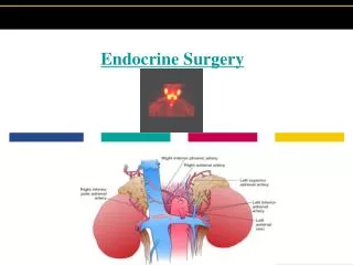
Problem: Inadvertent removal of parathyroid glands during endocrine surgery Iatrogenic injury common after procedures Resultant conditions of hypoparathyroidism and hypocalcemia can cause major health concerns Success primarily dependent upon surgeon experience No methods for visual distinction between similar tissues
Develop a real-time visualization system: • Optics lab have determined higher parathyroid autofluorescence compared to other neck tissues • Previous work undertaken to develop system for visualization system • Design Criteria: • System must be easy to use, requiring • little to no training • Function in real-time, with intuitive • information output • Easily integrated into surgical setting • Reduce time and error-rates of current • surgical practices
Objective: Idea: Employ relative thyroid and parathyroid auto-fluorescence to develop a real-time imaging system for use in endocrine surgery Find-R-Scope from previous Senior design project Drawbacks: Poor Image quality (low quality lenses) Surgical Considerations Unwieldy Configuration Limited penetration depth
Solution: IR Viewer Modification Method: - Diode laser (785nm) to emit excitation wavelength - Improve existing detector interface: - Alter handle configuration to allow reliable support (better integrate in surgical setting) - Add adapter to interface C-mount lenses - Determine optimal coupling of image into CCD - Write LabView VI to compile information - Employ false color for simple user interface - Test with sample tissue and in surgery
Considerations Imaging system must be able to clearly focus on entire surgical cut, approximately 6 inch diameter Sterile field surrounds patient: ~3 feet Surgeon needs visual indicator of parathyroid margin (focus <1 inch) within context of surgical field Imaging to be done no closer than 1 foot from patient Initial considerations are subject to change, input obtained from Dr. Phay and Dr. Broome of VUMC Endocrine surgery
Preliminary Results By removing front end of casing, further testing of lenses can be conducted
Preliminary Results At 3 feet from target, Navitar lens couple straight into imaging tube. Far (left) and near (right) focus images
Goals: - Determine if fixed zoom lens will provide higher resolution - Determine necessary excitation parameters - Improve necessary program interface - Find appropriate fluorescence standard for threshold creation - Verify system in vitro
Performance Indicators: Percentage of accurate identifications made when compared to histology and surgeon determination Time required for surgical procedure Cost of implementing system
Continuing Work: Redesign of lens coupling interface is underway (physical design of viewer) Researching fixed zoom lenses capable of video imaging necessary scale Upcoming: Developing software for fluorescence quantification Determine optimal filter configuration for desired image Determine Fluorescence standards or phantom creation