Download
1 / 72
720 likes | 902 Views
Radiologic Testing: What, When, & Why. Harry Colt, MD 7/20/09. Why This is an Important Topic. Radiology skills are underemphasized in medical school Radiology was a relatively weak part of our curriculum Most of our focus is on interpretation of X-rays

E N D
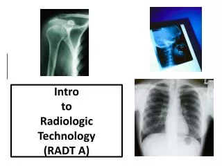
Radiologic Testing: What, When, & Why Harry Colt, MD 7/20/09
Why This is an Important Topic • Radiology skills are underemphasized in medical school • Radiology was a relatively weak part of our curriculum • Most of our focus is on interpretation of X-rays • Deciding what film to order is as important as interpreting the film • Residents now do a 2 week radiology rotation in year 3
Objective: • Participants will be able to identify appropriate • X-ray tests for many common clinical • conditions
Methods: • Brief orientation and review of the 7 main radiologic testing modalities • Case based approach
Seven Radiologic Modalities • plain films • Contrast Agents • CT scans • Ultrasound • Nuclear Imaging • Magnetic Resonance Imaging • PET Scan
Plain Films • image formed by attenuation of x-rays by the material that they are passing through • the denser the material, the greater the attenuation, the lighter the image will be • the four basic densities in order of increasing density: air, fat, water (blood, soft tissue), bone. It’s the contrast between these densities that delineates structures • plain films are 2D pictures of 3D structures, so multiple views generally needed
Contrast Agents • plain films are useful in situations where there is a natural contrast between body structures (e.g., heart & lung) • If no inherent contrast, contrast agents can help (esp. GI, urinary tract, and vasculature) • Disadvantages: • -5-10% have mild reaction: feel warm, metallic taste, etc • -0.1% have severe reaction: syncope, anaphylaxis, hypotension • -with low-osmolality agents, only 2% have reactions, but costs up to 10 times as much • -with IV contrast, increased risk of nephrotoxicity in patient with Cr≥1.5, particularly if diabetic
CT Scans • Rotating beam of x-rays that pass through patient and computer calculates absorption at thousands of points • Most organs (heart, kidney, liver, spleen, pancreas, etc.) are of uniform density and produce grey image on plain film. CT provides shades of grey. • Traditional CT: takes pictures like slices of loaf of bread
CT Scans cont’d • Spiral CT: pictures taken like paring of apple • Advantages of CT: • -differentiate structures of similar density • -view multiple structures simultaneously • Disadvantage • -many times the radiation of plain films • (see slide 5) • -generally need IV contrast with CT unless ruling out CNS bleed, ureteral “stone” protocol, or sinus views
Ultrasound • emits high frequency sound waves, assesses the strength and timing of returning echoes • us waves greatly reflected by air – soft tissue and bone – soft tissue interfaces, limiting its use in the chest and bones
Ultrasound cont’d • Advantages • -no radiation (safe in obstetrics) • -good for rapidly moving structures (e.g., heart) • Disadvantages • -limited in chest and bones • -operator dependent
Nuclear Imaging • Takes advantage of selective uptake of certain compounds in different organs of the body • These compounds can be labeled by radioactive isotopes • Their uptake can be recorded by a gamma camera that records radiation • Advantages: • -can obtain an image of function • Disadvantages: • -radiation (see slide 5) • -cost
MRI (Magnetic Resonance Imaging) • Applies magnetic field to the body. When field released, radio waves generated • Advantages: • -no ionizing radiation • -extraordinary views of CNS & stationary soft tissues • -contrast (Gadolinium) generally not needed unless MRA-neck, or MRI-head to rule out tumor • Disadvantages: • -inability to bring ferrous objects near magnet • -contraindications: pacer, defibrillator, aneurysm clips • -must hold still • -if gadolinium used, risk of nephrogenic systemic fibrosis in patients with renal failure
PET Scan • Allows imaging of structures based on their ability to concentrate specific molecules that have been labeled by positron emitting isotope • PET better than CT at differentiating benign from malignant lesions
Case #1 (Low Back Pain) 48 yo man presents with a 2 day history of severe low back pain radiating down posterolateral aspect of right leg to foot. Developed after gardening all day. No prior back problems. On exam: in obvious discomfort with movement. He has no neurologic deficits. Does he need imaging procedure? Why?
Case # 1 (Low Back Pain) The patient returns one week later with unchanged symptoms and exam. Does he need imaging procedure? If so, what type? Why?
Case # 1 (Low Back Pain) • Ninety plus percent of these patients recover spontaneously • Consider MRI at 6 weeks if not improving • Early plain films ($300) or MRI ($1600) indicated only if suspicion of fracture (significant recent trauma), infection, cancer, or progressive neurologic loss • Cont’d
Cont’d • Without these suspicions, early MRI results in increased frequency of surgical procedures, but no improved outcome • MRI> in asymptomatic individuals: • -52% with symmetric disc bulges • -27% with asymmetric disc bulging • -10% with disc extrusion • -75-80% of asymptomatic men over age 50 have disc bulging
Case #2 (Diabetic Foot Ulcer) 66 yo diabetic woman presents with 1 week history of 2cm ulceration on right foot. Does her foot need imaging? Why? Cont’d
Cont’d • If so, what technique? plain films, bone scan, or MRI? • Diabetic Foot Ulcers • in diabetic foot ulcers larger than 2cm2, 68% have osteomyelitis by bone biopsy and culture • Most have no sign of inflammation on exam
Case #2 (Diabetic Foot Ulcer)-cont’d • plain Films ($271) • Can identify soft tissue swelling, bone destruction, periosteal elevation • insensitive for acute Osteomyelitis. 2-3 weeks usually needed to see bony changes • Even after 3 weeks, sensitivity approaches 60-80% • Cont’d
Cont’d • 3 Phase Bone Scan ($900) • Technetium bound to phosphorus, and accumulates in areas of increased osteoblastic activity • 3 phases • -1st phase: immediate – reflects flow • -2nd phase: 15 min. – reflects blood pooling • -3rd phase: 4 hours – bone imaging • cont’d
Cont’d • 3 Phase Bone Scan • Cellulitis has increased activity in phases 1 and 2 • Osteomyelitis has intense uptake in all 3 phases • Often times positive in acute osteo by 3 days • Imaging procedure of choice for acute osteomyelitis • Cont’d
Case #2 (Diabetic Foot Ulcer)-cont’d • MRI ($1500) • can be very useful in infected diabetic foot • Sensitivity 95% • Imaging test of choice for chronic osteo
Case #3 (Abdominal Pain) 54 yo woman presents with 3 day history of upper abdominal pain, nausea, and occasional vomiting. On exam: Temp. 100o, tender in RUQ,. Labs: wbc 14.8, normal LFTs, lipase, and amylase. What is the most likely diagnosis? What is your imaging procedure of choice? Why?
Case #3(cholecystitis) • Ultrasound ($698) • can identify stigmata of cholecystitis: • -gallstones • -gallbladder wall thickening (>4-5 mm) • -gallbladder wall edema (double wall sign) • -sonographic Murphy’s sign • For cholecystitis: sensitivity 88%, specificity 80% • Can miss very small stones (<3mm)
Case #3 (Cholecystitis)-cont’d • Cholescintigraphy (HIDA scan) ($1200) • Use technetium labeled hepatic iminodiacetic acid • Injected IV, taken up by hepatocytes and excreted in bile • If the cystic duct is patent, it will enter gallbladder • Test is positive (abnormal) if gallbladder not visualized, usually due to cystic duct obstruction from edema from cholecystitis or stone • Sensitivity 97%, specificity 90%
Case #4 (Hip Injury) 88 yo male presents with hip pain after fall last night. On exam: complains of pain with any movement of hip. Initial hip films are inconclusive for fracture. What is the imaging test of choice when hip fracture is suspected, but plain films are negative?
Case #4 (Hip Injury)-cont’d • MRI ($1500) is study of choice • Bone scan ($1100) indicated for suspected fracture when MRI not available or contraindicated • CT ($1200)
Case #5 (Diverticulitis) 77 yo man presents with LLQ pain and nausea for 2 days. On exam has temp of 101o, LLQ tenderness. What is imaging procedure of choice?
Case #5 (Diverticulitis) • plain films? ($475) No • abdominal films usually only helpful when you suspect obstruction or significant perforation • CT? ($1631) Yes • Helical CT with contrast: sensitivity 97% for diverticulitis features include: increased soft tissue density secondary to inflammation (“greying” of fat), colonic diverticula, bowel wall thickening, soft tissue masses • Contrast Enema? ($900) In rare cases • Would use water soluble contrast given risk of perforation.
Case #5A (Diverticulitis) – same patient 77 yo man with diabetes with LLQ pain and nausea for 2 days. On exam has temp of 101o, LLQ tenderness. Creat 1.8. What is the imaging procedure of choice?
Discuss with radiologist: CT without IV contrast vs CT with contrast vs MRI without contrast or US If opt for CT with contrast, patient needs: -ISO osmolal agent -avoid volume depletion and NSAIDS -if no contraindictions, IV isotonic fluids -consider acetylcystine
Case #6 (Sinusitis) 34 yo female presents with 3 week history of nasal congestion & maxillary tenderness. Believes she has recurrence of sinusitis. Does she need imaging?
Case #6 (Sinusitis) • If patient believed to have sinusitis, would treat without imaging. • If fails treatments, then CT is imaging procedure of choice. • plain sinus films (3v) ($372) have low sensitivity • CT ($960) much more sensitive, but gives false positives. 27/31 false positives in 1 study of patients with cold. Don’t order early in course of illness, you will only generate unhelpful information.
Case #7 (Urolithiasis) 64 yo man presents with 1 day history of severe left flank pain. Never had similar symptoms previously. On exam, his abdomen is nontender, no prominent abdominal pulsation. Urine shows microscopic hematuria. You suspect ureteral stone. What is imaging procedure of choice?