Download
1 / 27
270 likes | 291 Views
Learn about the structure of the plasma membrane, including the phospholipid bilayer and embedded proteins. Discover how membrane fluidity and cholesterol affect membrane function.

E N D
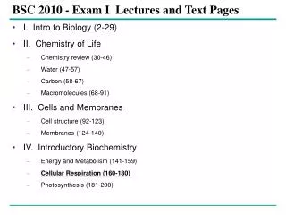
BSC 2010 - Exam I Lectures and Text Pages • I. Intro to Biology (2-29) • II. Chemistry of Life • Chemistry review (30-46) • Water (47-57) • Carbon (58-67) • Macromolecules (68-91) • III. Cells and Membranes • Cell structure (92-123) • Membranes (124-140) • IV. Introductory Biochemistry • Energy and Metabolism (141-159) • Cellular Respiration (160-180) • Photosynthesis (181-200)
The Plasma Membrane – Structure and Function • Is the boundary that separates the living cell from its nonliving surroundings • The basic structure is a phospholipid bilayer.
Phospholipids • Are the most abundant lipid in the plasma membrane • Are amphipathic, containing both hydrophobic and hydrophilic regions
Figure 7.1 Fluid-Mosaic Model • The model states that: The plasma membrane is a phospholipid bilayer with many things floating in it (also amphipathic molecules).
Fluid-Mosaic Model • Most of the mosaic molecules are proteins, including many for transport. 1. Channels- gates that can open/close, mainly for ion transport 2. Carriers - pick up a molecule and rotate into the cell. 3. Receptors- mainly for hormones. When the hormone binds to the receptor, the receptor protein changes shape and stimulates a secondary messenger within the cell. 4. Cell-cell recognition- mainly for self-identification. e.g. H-Y antigen present on all cells of males, makes organ donation not work from male to female. e.g. Blood types (ABO) and the Rh factor. Recognition proteins are apparently not functional in fetuses or newborns.
Fluid-Mosaic Model • Carbohydrates can also be part of self-recognition, or for membrane-membrane interactions (e.g. tissue glue that holds cells together). This is not the ECM, but is part of the ECM. There's more to it than that. Look at the end of the chapter. • Be sure to review the structure of the membrane and predict what types of molecules can and can't pass through the phospholipid bilayer.
WATER Hydrophilic head Hydrophobic tail WATER Figure 7.2 Membrane Models • Membranes have been chemically analyzed, and they have in fact been found to be composed of proteins and lipids. • Scientists studying the plasma membrane reasoned that it must be a phospholipid bilayer.
Davson-Danielli sandwich model of membrane structure • Stated (1935) that the membrane was made up of a phospholipid bilayer sandwiched between two protein layers • Was supported by electron microscope pictures of membranes. • But predicted that all membranes were of the same makeup, which they are not. • And does not fit with the fact that many membrane proteins are amphipathic and have hydrophobic regions (are not water-soluble).
Hydrophobic region of protein Phospholipid bilayer Figure 7.3 Hydrophobic region of protein In 1972, Singer and Nicolson • Proposed that membrane proteins are dispersed and individually inserted into the phospholipid bilayer
A cell membrane can be split into its two layers, revealing the ultrastructure of the membrane’s interior. APPLICATION TECHNIQUE Extracellular layer Proteins Knife Plasma membrane Cytoplasmic layer These SEMs show membrane proteins (the “bumps”) in the two layers, demonstrating that proteins are embedded in the phospholipid bilayer. RESULTS Freeze-fracture studies of the plasma membrane • Supported the fluid mosaic model of membrane structure A cell is frozen and fractured with a knife. The fracture plane often follows the hydrophobic interior of a membrane, splitting the phospholipid bilayer into two separated layers. The membrane proteins go wholly with one of the layers. Figure 7.4 Extracellular layer Cytoplasmic layer
Lateral movement (~107 times per second) Flip-flop (~ once per month) (a) Movement of phospholipids Figure 7.5 A The Fluidity of Membranes • Phospholipids in the plasma membrane • Can move within the bilayer • Fluidity affects transport and the functioning of proteins.
Membrane proteins EXPERIMENT Researchers labeled the plasma membrane proteins of a mouse cell and a human cell with two different markers and fused the cells. Using a microscope, they observed the markers on the hybrid cell. RESULTS Mouse cell Mixed proteins after 1 hour Human cell Hybrid cell + CONCLUSION The mixing of the mouse and human membrane proteins indicates that at least some membrane proteins move sideways within the plane of the plasma membrane. Figure 7.6 Proteins in the plasma membrane • Can drift within the bilayer
Fluid Viscous Unsaturated hydrocarbon tails with kinks Saturated hydro- Carbon tails (b) Membrane fluidity Figure 7.5 B The type of hydrocarbon tails in phospholipids • Affects the fluidity of the plasma membrane
Cholesterol Figure 7.5 (c) Cholesterol within the animal cell membrane The steroid cholesterol • Has different effects on membrane fluidity at different temperatures • Reduces phospholipid movement (fluidity) • But, hinders solidification at low temperatures
Glycoprotein Carbohydrate Glycolipid EXTRACELLULAR SIDE OF MEMBRANE Microfilaments of cytoskeleton Peripheral protein Cholesterol Integral protein CYTOPLASMIC SIDE OF MEMBRANE Figure 7.7 Membrane Proteins and Their Functions • A membrane • Is a collage of different proteins embedded in the fluid matrix of the lipid bilayer Fibers of extracellular matrix (ECM)
N-terminus C-terminus CYTOPLASMIC SIDE a Helix Figure 7.8 Integral proteins • Penetrate the hydrophobic core of the lipid bilayer • Are often transmembrane proteins, completely spanning the membrane EXTRACELLULAR SIDE
EXTRACELLULAR SIDE OF MEMBRANE Peripheral protein Integral protein CYTOPLASMIC SIDE OF MEMBRANE Figure 7.7 Peripheral proteins • Are appendages loosely bound to the surface of the membrane
Transport. (left) A protein that spans the membrane may provide a hydrophilic channel across the membrane that is selective for a particular solute. (right) Other transport proteins shuttle a substance from one side to the other by changing shape. Some of these proteins hydrolyze ATP as an energy source to actively pump substances across the membrane. (a) ATP (b) Enzymatic activity. A protein built into the membrane may be an enzyme with its active site exposed to substances in the adjacent solution. In some cases, several enzymes in a membrane are organized as a team that carries out sequential steps of a metabolic pathway. Enzymes Signal transduction. A membrane protein may have a binding site with a specific shape that fits the shape of a chemical messenger, such as a hormone. The external messenger (signal) may cause a conformational change in the protein (receptor) that relays the message to the inside of the cell. (c) Signal Receptor Figure 7.9 Six major functions of membrane proteins
(d) Cell-cell recognition. Some glyco-proteins serve as identification tags that are specifically recognized by other cells. Glyco- protein (e) Intercellular joining. Membrane proteins of adjacent cells may hook together in various kinds of junctions, such as gap junctions or tight junctions (see Figure 6.31). (f) Attachment to the cytoskeleton and extracellular matrix (ECM). Microfilaments or other elements of the cytoskeleton may be bonded to membrane proteins, a function that helps maintain cell shape and stabilizes the location of certain membrane proteins. Proteins that adhere to the ECM can coordinate extracellular and intracellular changes (see Figure 6.29). Figure 7.9
The Role of Membrane Carbohydrates in Cell-Cell Recognition • Cell-cell recognition • Is a cell’s ability to distinguish one type of neighboring cell from another • Membrane carbohydrates • Interact with the surface molecules of other cells, facilitating cell-cell recognition
Synthesis and Sidedness of Membranes • Membranes have distinct inside and outside faces • This affects the movement of proteins synthesized in the endomembrane system
1 Transmembrane glycoproteins Secretory protein Glycolipid 2 Golgi apparatus Vesicle 3 Plasma membrane: Cytoplasmic face 4 Extracellular face Transmembrane glycoprotein Secreted protein Membrane glycolipid Figure 7.10 Membrane proteins and lipids • Are synthesized in the ER and Golgi apparatus ER
Selective Permeability • Membrane structure results in selective permeability • A cell must exchange materials with its surroundings, a process controlled by the plasma membrane
A Selectively Permeable Barrier • The plasma membrane exhibits selective permeability. It controls what substances enter and leave the cell. • It allows some substances to cross more easily than others
The Permeability of the Lipid Bilayer • Hydrophobic molecules • Are lipid soluble and can pass through the membrane rapidly • Hydrophilic substances • Do not cross the membrane rapidly • Includes polar molecules and ions
A Selectively Permeable Barrier • Things that pass easily through the bilayer: 1. small non-polar molecules (hydrocarbons, O2, N2) 2. small polar uncharged molecules (H2O, CO2, glycerol, urea) • Things that don’t pass easily through the bilayer: (Require transport) 1. large polar molecules (glucose) 2. ions (H+, Na+, Cl-, Mg++, PO42-)
Transport Proteins • Transport proteins • Allow passage of hydrophilic substances across the membrane • Transport may be passive by diffusion, which follows the concentration gradient of the molecules and requires no expenditure of energy by the cell. • Transport may be active, against the concentration gradient, requiring an energy source, generally ATP.