Download

1 / 66
660 likes | 1.21k Views
Functions Of The Spinal CordConduction ? contains bundles of nerves that conduct information up and down the cord ? enables sensory information to reach the brain, motor commands to reach the effectors, and input received at one level of the cord to affect output from another levelLocomotion ? wal

E N D
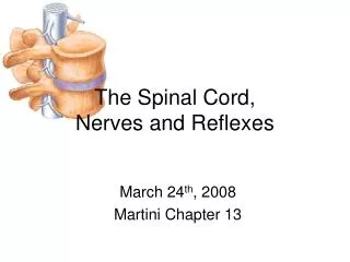
1. The Spinal Cord,Spinal Nerves,and Somatic Reflexes Chapter 13
2. Functions Of The Spinal Cord
Conduction � contains bundles of nerves that conduct information up and down the cord � enables sensory information to reach the brain, motor commands to reach the effectors, and input received at one level of the cord to affect output from another level
Locomotion � walking
Reflexes � involve the brain, spinal cord and peripheral nerves
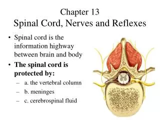
3. Spinal Cord � begins at the foramen magnum of the skull and passes through the vertebral canal as far as the inferior margin of the first lumbar vertebra � occupies only the upper two thirds of the vertebral canal � gives rise to 31 pairs of spinal nerves that pass through the intervertebral foramina
Segments � the part supplied by each pair of spinal nerves
Longitudinal grooves � ventral median fissure and dorsal median sulcus
4. Spinal cord divisions � cervical, thoracic, lumbar and sacral � named for the level from which the spinal nerves emerge
Cervical enlargement � area in the inferior cervical cord where the cord gives rise to nerves of the upper limbs
Lumbar enlargement � an area in the lumbosacral cord where the cord gives rise to nerves in the pelvic region and the lower limbs
5. Medullary cone � inferior to the lumbar enlargement the cord tapers to a point called the medullary cone
Cauda equina � the lumbar enlargement and the medullary cone give rise to a bundle of nerve roots that occupy the vertebral canal from L2 � S5
8. Meninges of the spinal cord � three fibrous connective tissue membranes that separate the soft tissue of the central nervous system from the bones of the vertebrae and the skull
Dura mater � forms a loose fitting sleeve (dural sheath) around the spinal cord � space between the sheath and the vertebral bones is the epidural space
Arachnoid mater � membrane of simple squamous epithelium that adheres to the inside of the dura � gap between the arachnoid mater and the pia mater is called the arachnoid space and is filled with cerebsrospinal fluid
9. Pia mater � closely follows the contours of the spinal cord
Terminal filum � pia mater extends beyond the medullary cone as a fibrous strand that forms part of the coccygeal ligament which anchors the cord to the coccygeal vertebrae
Denticulate ligaments � extensions of the pia mater through the arachnoid to the dura anchoring the cord
12. Gray matter of the spinal cord � contains somas, dendrites, and proximal parts of axons � site of synaptic contact between neurons � very little myelin
Dorsal horns � dorsal root of the spinal nerve carrying sensory nerve fibers enters the dorsal horn of the cord and synapse with interneurons there � these neurons are especially numerous in the cervical and lumbar enlargements
13. Ventral horns � contain the large somas of the somatic motor neurons � axons from these neurons exit by way of the ventral root of the spinal nerve and lead to the skeletal muscle
Lateral horns � visible on each side of the gray matter from cord segments T2 through L1 � contains neurons of the sympathetic nervous system which send their axons out of the cord by way of the ventral root along with the somatic efferent fibers
14. White matter � bright appearance � abundance of myelin � composed of bundles of axons called tracts that carry signals from one part of the CNS to another � surrounds the gray matter
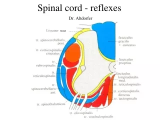
Three pairs of columns or funiculi � dorsal, lateral and ventral columns � each column consists of subdivisions called tracts or faciculi
15. Spinal Tracts
Ascending tracts � carry sensory information up the cord
Descending tracts � carry motor impulses down
Decussation � when tracts cross over from one side of the body to the other
Contralateral � when the origin and destination of a tract are on opposite sides of the body
Ipsilateral � when a tract does not decussate and the origin and destination of its fibers are on the same side of the body
16. Ascending tracts � carry sensory signals up the spinal cord � sensory signals typically travel across three neurons from their origin to their destination in the sensory areas of the brain
First order neurons � detects a signal and transmits it to the spinal cord or brainstem
Second order neuron � carries the signal to the thalamus
Third order neuron � carries the signal to the sensory region of the cerebral cortex
17. Ascending Tracts
Gracile fasciculus
Cuneate fasciculus
Spinothalamic
Spinoreticular
Dorsal and ventricular spinocerebellar
18. Descending tracts � carry motor signals down the brainstem and spinal cord � typically involve two neurons
Upper motor neuron � begins with a soma in the cerebral cortex or brainstem and has an axon that terminates on a lower motor neuron in the brainstem or the spinal cord
Lower motor neuron � the axon of the lower motor neuron then leads the rest of the way to the muscle or other target organ
19. Corticospinal tract � lateral and ventral
Tectospinal tract
Lateral and medial reticulospinal tract
Lateral and medial vestibulospinal tract
25. Anatomy of Nerves and Ganglia
Nerve is a cord composed of nerve fibers (axons) bound together by connective tissue
Nerve fibers in the PNS are ensheathed in Schwann cells which form a myelin sheath around the axon with a neurilemma � surrounded by basal lamina and then endoneurium
Nerves bundled in fasicles surrounded by perineurium
Several fasicles are bundled together and surrounded by an epineurium
27. Mixed nerve � consists of both sensory and motor fibers � transmits signals in two directions � any one fiber only transmits in one direction
Sensory nerves composed entirely of sensory axons are less common � olfactory and optic nerves
Motor nerves carry only motor fibers
Ganglion � a cluster of cell bodies outside the CNS � enveloped in an epineurium continuous with that of the soma � among the somas are nerve fibers leading into and out of the ganglion
30. Spinal Nerves
31 pairs of spinal nerves � 8 cervical (C1 - C8), 12 thoracic (T1 � T12), 5 lumbar (L1 � L5) , 5 sacral (S1 � S5), and 1 coccygeal
Proximal branches � each spinal nerve has two points of attachment to the spinal cord � dorsally a branch of the spinal nerve called the dorsal root divides into six to eight rootlets that enter the spinal cord � distal to the rootlets is a swelling called the dorsal root ganglion which contains somas of unipolar afferent neurons
31. Ventrally another row of six to eight rootlets leave the spinal cord and converge to form the ventral root
The dorsal and ventral roots merge, penetrate the dural sac , enter the intervertebral foramen and form the spinal nerve proper
Spinal nerves are mixed nerves with afferent sensory and efferent motor signals �
Afferent nerves approach the cord by way of the dorsal root and enter the dorsal horn of the gray matter
32. Efferent signals begin at the somas of motor neurons in the ventral horn and leave the spinal cord via the ventral root
Some viruses invade the central nervous system by way of these roots
33. Roots that arise from L2 to C0 form the cauda equina