Download
1 / 62
700 likes | 1.37k Views
Learn about the impact of device therapy on heart failure, including risks with right ventricular pacing, benefits of biventricular pacing, and advancements in implantable defibrillators. Discover how these technologies are improving patient outcomes.

E N D
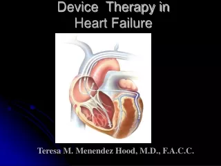
Device Therapy in Heart Failure Teresa M. Menendez Hood, M.D., F.A.C.C.
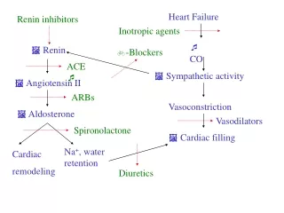
Heart Failure • Up to 30 % of HF patients have an IVCD (80% with a LBBB) which has been linked to increases in mortality and morbidity. • HF is the leading cause of hospitalizations in the US and uses up 5% of the health care costs • 2% of the population and 6% of the population >65 • Prevalence is on the rise. Annual Incidence Heart Failure Prevalence Annual Mortality 5.0 million 400,000 250,000 U.S.
NYHA Class-evaluates the disability imposed on the patient who already has structural heart disease Class I Asymptomatic heart failureejection fraction (EF) <40% Class II Mild symptomatic heart failure with ordinary exertion Class III Moderate symptomatic heart failure with less than ordinary exertion Class IV Symptomatic heart failure at rest
Leading Causes of Death in the U.S. Septicemia You must combine deaths from all cancers to outnumber the deaths from SCA each year. Nephritis Alzheimer’s Disease Influenza/pneumonia Diabetes Accidents/injuries Chronic lower respiratory diseases Cerebrovascular disease Other cardiac causes Sudden cardiac arrest (SCA) All other causes All cancers 0% 5% 10% 15% 20% 25% National Vital Statistics Report. Oct. 12, 2001;49(11). MMWR. State-specific mortality from sudden cardiac death – US 1999. Feb 15, 2002;51:123-126.
SCD Rates in CHF Patients with LV Dysfunction 45 months 13 months 41.4 months 27 months 12 months 16 months 6 months SCD accounts for ~50% of the total deaths.
SCD in Heart Failure • QRS duration is an independent predictor of mortality (>140 ms) • Other factors are: age, creatinine, EF, and HR QRS 100% Duration (msec) <90 90% 90 - - 120 Cumulative Survival 80% 120 - - 170 170 - - 220 70% >220 60% 0 60 120 180 240 300 360 Days .
SCD in Heart Failure • Degree of SCD risk by class • Mortality in NYHA class II is 5 to 15% • 50 to 80% of the deaths are Sudden • Mortality in NYHA class III is 20 to 50% • Up to 50% of the deaths are Sudden • Mortality in NYHA class IV is 30 to 70% • 5 to 30% of deaths are Sudden (more deaths from pump failure)
Right Ventricular Pacing • RV apex pacing is harmful in patients with LV dysfunction. Became evident in multiple pacer and ICD trials that it increases HF by producing a “paced” LBBB. • Abnormal LV activation • Reduced stroke volume
Detrimental RV pacing • MADIT II (2002) had a survival benefit with the ICD but in a subgroup analysis, there was an increase in heart failure morbidity (more hospitalizations) felt due to forced RV pacing compared to controls in which no pacing was present.
(p= 0.09) N= 490 N= 742 MADIT II: ComplicationsNew or Worsening HF • RV pacing causes ventricular dysynchrony and may lead to worsening HF. • Intrinsic ventricular activation is better for ICD patients with left ventricular dysfunction who do not “need” pacing. • <10% of ICD patients have a Class I pacing indication at the time of implant…they do not NEED pacing. • Physicians, when appropriate, should consider programming of ICDs to avoid frequent RV pacing.
DAVID — Dual Chamber and VVI Implantable Defibrillator Trial : 2002 • ICD indication but no indication for a pacemaker • EF < 40% • DDDR @ 70BPM versus VVI 40 BPM
Search AV Extension and Managed Ventricular Pacing for Promoting Atrioventricular Conduction (SAVE PACe) Trial : 2007 • 1065 patients with sinus-node disease, intact AV conduction and normal QRS interval • Randomized to conventional dual-chamber pacing (n=535) or dual-chamber minimal ventricular pacing (n=530) ― study tests new pacing algorithm that avoids ventricular pacing except during periods of high-grade AV block • With dual-chamber pacing, ↓ frequency RV pacing (9.1% vs. 99%; p<0.001) and 40% relative risk ↓ in incidence of persistent AF
TheConcept • In most patients with an IVCD (QRS > 130 ms) , the presence of atrial-biventricular (RV + LV) pacing will provide early stimulation to an otherwise late segment of electrical activation in the LV. • This should translate into an increase in the EF, decrease of the LV dimension, improvement in the QOL and NYHA class. • This may translate into an decrease in CHF exacerbations , hospitalizations and a decrease in mortality.
The Proof • 1994 –1997: Mechanistic and both short and longer term observational studies. Studies initially used epicardial leads placed by thoracotomy or thorascope. • The first BiV pacer was implanted in 1994 • 1998 –1999: Randomized, controlled studies to assess exercise capacity, functional status, and quality of life. • There was development of transvenous leads via the coronary sinus in to get to the LV. Cohen TJ, Klein J. J Inva2002;14:48-53.
The Proof • 2000 – 2006: Randomized, controlled trials to assess combined mortality and CHF hospitalization. Also evaluated the combined benefit of ICD’s with CRT. • 2006-2008: Trials to identify patients who will benefit from CRT. This uses echocardiographic markers of dyssynchrony and the QRS measurement. • 20% of patients do not respond to therapy in clinical trials with a wide QRS and 50% patients with a narrow QRS/CHF have dyssynchrony on echo and may benefit from this therapy. • If the QRS is < 150 ms, then the chance of responding to BiVP is ~5%. It will be in this patient group of QRS of 120-150 ms where preselection of responders would be most valuable.
The Cardiac Resynchronization Clinical Trials PATH-CHF, MUSTIC, MIRACLE, COMPANION, and CARE-HF* *This is not a complete list of all the CRT trials and the dates given are when the trial results were published.
Cumulative Enrollment in Cardiac Resynchronization Randomized Trials
PATH-CHF: 1999 Pacing Therapy for Congestive Heart Failure • This was the first multicenter trial and used the standard endocardial RV lead and an epicardial LV lead via thoracotomy or thorascope • Single blinded RCT • 53 centers in Europe • 41 patients
PATH-CHF • Primary endpoints • Peak VO2 • Six-minute walk distance • Secondary endpoints • Minnesota Living with Heart Failure score (QOL) • NYHA class • EF • Trend towards decrease in Hospitalizations • Acute hemodynamic testing revealed that the lateral and posterolateral walls were the best target sites. • The best responders were those with QRS>150 , long PR and dP/dt < 700 mm Hg/s
MUSTIC: 2001Multicenter Stimulation in CM • European study with 67 patients • QRS>150, CHF, EF <35% • BiVP versus backup VVI pacing at 40 BPM • Increase in 6 minute walk time , QOL and Peak VO2 with BiVP and persisted for up to 12 months • 60% decrease in CHF hospitalizations • First to use endocardial LV leads via the CS • No significant change in mortality, but a trend towards an improvement. • Acute hemodynamic studies showed the mid lateral wall to be the best site
MIRACLE:2002Multi-center In Sync Randomized Clinical Evaluation Trial • Double blinded RCT • FirstUS trial • Class 3 or 4, on OPT, QRS >130 ms, EF<35% • Enrollment of 453 patients
NYHA class III-IV LVEDD > 60 mm QRS > 130 ms Stable 3 month regimen of beta-blocker/ACE inhibitor EF < 35% Randomization CRT on CRT off 1- and 3-month follow-up 1- and 3-month follow-up 6-month follow-up 6-month follow-up CRT on Long-term follow-up MIRACLE
P < 0.001 67% 60% 40% Proportion 39% 34% 27% 20% 17% 16% 0% Improved No Change Worsened Control N=225 CRT N=228 MIRACLE Nonresponders: older, ischemic CM, no MR, QRS<150 Responders: had shorter duration on CHF and longer QRS>155
MIRACLE • There was a decrease in hospitalizations of 50% at 6 months and a trend towards a decrease in mortality. • All other primary and secondary endpoints were met: 6 minute walk time, peak Vo2, QOL, EF , NYHA class, LVEDD Magnitude of improvement not influenced by degree of QRS shortening with BiVP (average in all was –20msec)
FDA Approval • The first CRT device was approved by the FDA in September 2001 . • The first CRT with an ICD was approved by the FDA in May 2002 .
The Primary ICD Prevention Trials • MADIT 11996 required a positive EP study;ischemics • MUSTT1999 required a positive EP study; ischemics; EF<40% • MADIT 22002 prior MI (ischemic cardiomyopathy) and EF<30% (no EP study required) ;60% had CHF and 50% had QRS > 120 ms; resulted in a 31% decrease risk of death and halted prematurely due to the positive effect of the ICD: resulted in the FDA approving the ICD for primary prevention this patient population, but only those with a QRS > 120 ms.
The Primary ICD Prevention Trials • SCD-Heft -2005 The SCD-Heft trial resulted in FDA approval of the ICD January 2005 in patients with CHF and EF<35 % that included both ischemic and nonischemic cardiomyopathy for primary prevention without a positive EP study or ventricular ectopy . No QRS cutoff was required.
1 • OPT 2 + • OPT • CRT Randomization 2 • OPT • CRT-D + COMPANION:2004 Comparison of Medical Therapy, Pacing and Defibrillation in Heart Failure
COMPANION • Enrolled 1520 patients class 3 and 4, QRS >120ms • Primary endpoint: death or hospitalization for any cause • CRT met the primary endpoints and the CRT +/- ICD significantly reduces mortality • This was the first to show mortality benefit from CRT alone • Showed that patients with CRT also benefit from ICD therapy • OPT had SCD in 36%,23% in CRT and 3% in CRT+ICD
COMPANION • CRT arm had 20% reduction in mortality and hospitalization over OPT arm but it was not statistically significant • Significant reduction in CRT-ICD arm of 40% for mortality over OPT arm (19% in OPT and 11% in CRT-ICD group) • Study was halted prematurely due to its positive benefit. • Mean follow up was 16 months
CARE-HFCArdiac REsynchronization in Heart Failure 2005 • The effect of cardiac resynchronization on morbidity and mortality in heart failure in 813 patients in Europe ( prospective multicenter RCT) with completed enrollment by 2002 • Large patient size and length of trial (average follow up of 29 months) allowed ability to asses effects of CRT • Looked at CRT alone (no ICD) • Patients with class 3 or 4, EF < 35%, QRS >120 ms • There was a 37% reduced mortality or first hospitalization for a cardiac cause compared to OPT
CARE-HF • All endpoints were met : EF, NYHA, QOL, BNP, Echo and hemodynamic parameters • 33% of the deaths in the CRT group were due to SCD • For every 9 devices, one death and 3 hospitalizations were prevented • Echo criteria in patients with QRS 120-149ms to look for dyssynchrony (had to have 2 of 3)…the “gray area group” • Aortic pre-ejection delay of > 140 ms ( onset of QRS to Aortic ejection) • Interventricular mechanical delay of >40 ms ( RV-LV) • Delayed activation of the postero-lateral LV wall (>50ms)
1.00 HR 0.63 (95% CI 0.51 to 0.77) 0.75 0.50 Event-free Survival P < .0001 Medical : 224 ptsTherapy (55 %) 0.25 0.00 0 500 1000 1500 Days Number at risk 409 323 273 166 68 7 CRT 404 292 232 118 48 3 Medical Therapy Primary Endpoint(All-cause Mortality or Unplanned Hosp. for Major CVS Event) CRT : 159 pts (39%)
Conclusions • Conclusive results from CARE-HF demonstrate that CRT should be considered as part of routine therapy for patients with moderate to severe HF due to LVSD with evidence (ECG supported by Echo) of cardiac dyssynchrony to*: • Improve cardiac function and efficiency • Improve symptoms and QoL • Reduce morbidity • Prolong survival • These benefits are in addition to those of optimal pharmacological therapy (OPT)
The Resynchronization Therapy in Normal QRS (RethinQ) Study2007
Background Currently, indications for cardiac resynchronization therapy (CRT) include QRS duration > 120ms, LVEF < 35% and NYHA Class III-IV. 20-30% of patients do not respond to CRT despite application of established selection criteria. Patients with normal conduction or a slightly prolonged QRS duration also exhibit mechanical abnormalities due to intraventricular dyssynchrony. Myocardial Tissue Doppler Imaging (TDI) allows both the velocity and timing of regional longitudinal motion to be measured. LV dyssynchrony may also be useful in predicting the benefit of CRT before implantation of the pulse generator.
Hypothesis We hypothesized that patients with NYHA class III, left ventricular ejection fraction less than or equal to 35%, narrow QRS duration < 130 ms, and evidence of mechanical dyssynchrony on echocardiography may benefit from cardiac resynchronization therapy.
Echo Criteria for LV Dyssynchrony Mechanical dyssynchrony considered present if either • M-Mode - Septal posterior wall mechanical delay (SPWMD) ≥ 130 ms OR • Tissue Doppler Imaging (TDI) of the basal ventricular segments in apical 4/2/3 chamber views - Septal to lateral delay ≥ 65ms OR - Antero-septal to posterior delay ≥ 65ms
Summary:RethinQ This prospective, multi-center, randomized trial was designed to evaluate the effectiveness of CRT therapy in a HF population with narrow QRS duration and evidence of mechanical dyssynchrony. There was no statistical significant difference in the change in Peak VO2 between the treatment and control group during cardiopulmonary exercise testing. No improvement in other objective parameters including 6-minute walk test, LV reverse remodeling, and secondary endpoint - quality of life score .
Conclusion:RethinQ CRT did not improve Peak VO2 during exercise in patients with NYHA Class III heart failure, QRS duration <130ms, EF ≤ 35% and mechanical dyssynchrony as specified in this trial. While there was a statistically significant improvement of NYHA class, a secondary endpoint, there was no improvement in quality-of-life, 6-minute walking test, or echocardiographic measures of reverse LV remodeling A subgroup of patients with QRS duration between 120 ms and 130 ms demonstrated an improvement from CRT, however patients with QRS duration < 120 ms did not demonstrate improvement The subgroup of patients stratified on the basis of cardiomyopathy etiology did not demonstrate an improvement in peak VO2.
PROSPECT TRIAL 5/2008 • Predictors of response to CRT • 53 centers worldwide, 426 patients • Patients had standard CRT indications (OMT, EF < 35%, Class III-IV, QRS > 130) • 12 ECHO parameters of dyssynchrony • 69% of patients clinically improved and 56% showed a decrease in LVESV of >15% • No single ECHO measure of dyssynchrony could help select responders to CRT
BASE Anterior Posterior APEX RAO is best to distinguish BASE position from APEX
ANTERIOR Anterior LAO is best to distinguish LATERAL position from SEPTAL LATERAL SEPTAL Posterior Lateral INFERIOR
The 3 levels of Dyssynchrony • Intraventricular dyssynchrony is best treated by placing the LV lead in the best anatomiclocation-usually the lateral or posterolateral (proven my multiple studies). Get the LV working. • Interventricular dyssynchrony is dealt with by adjusting the V-V interval. Get the RV and the LV to work together. • A-V dyssynchrony is dealt with by adjusting the A-V interval. Get the atria and the ventricles working together.
Posterolateral or Lateral walls are the best with LBBB where the septum contracts first and then the lateral wall last. Paced at most mechanically delayed LV site Paced at any other LV site 0 10% P=0.04 -5 -9.2 8% 9% -10 6% Improvement -15 4% -20 2% -25 -28.4 P=0.04 2% -30 0% Change in LV End-systolic Volume [ml] Change in LVEF [%]
CRT and Tissue Doppler Imaging -a measure of intraventricular delay • Measures dyssynchronous (delayed) contraction patterns @ different areas of the ventricle • Measure from the onset of the QRS to the peak systolic shortening of that segment • Defined as a segment with > 50 ms delay: this indicates intraventricular delay or asynchrony by ECHO criteria • Colors: green-yellow-red (the longest delay of >300 ms)
V-V Timing: synchronize the RV and the LV • The best V-V setting by measuring the RVOT and LVOT via PW Doppler • V-V above > 40 ms is considered abnormal • In normals, the RV will contract before the LV in the heart by -20 ms • LV and RV have different outputs in the newer devices that allow sequential instead of simultaneous delivery of output and thus allow for this to be programmable.